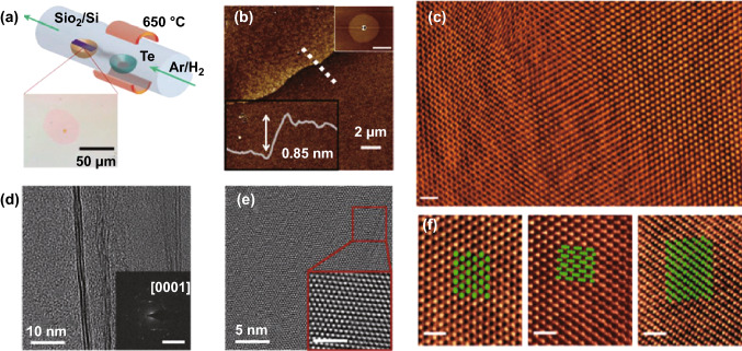
Fig. 2.
Ultrathin Te flakes synthesized by PVD. a Schematic of the experimental setup. b Atomic force microscopy (AFM) image of the edge of a Te flake including a profile taken along the dotted line showing the thickness of the flake. c High-angle annular dark-field scanning transmission electron microscopy (HAADF–STEM) image of Te flakes showing the large-scale uniformity. d, e TEM images of Te flakes showing their structure measured by electron diffraction (inset of d). f Atomically resolved HAADF–STEM images of the three Te polymorphs. Adapted with permission from [66]. Copyright 2019, WILEY–VCH

