Fig. 4.
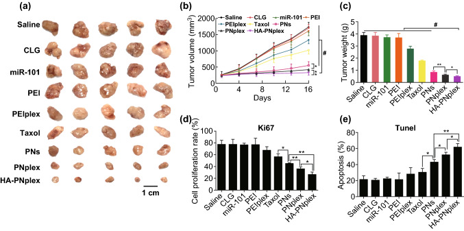
Anti-tumor efficiency. Different formulations (0.2 mL) were injected into MCF-7 tumor-bearing nude Balb/C mice via tail vein every 3 days at a PTX dose of 10 mg kg−1 and miR-101 dose of 1 mg kg−1, according to the animal’s body weight. On day 16 at the end of the experiment, the tumor tissues were collected for further examination. a Representative images of the tumors collected from mice at the end of the experiment. b Tumor growth curves (n = 6, *P < 0.05, **P < 0.01 and #P < 0.001). c Tumor weight after treatment with the various formulations (n = 6, *P < 0.05, **P < 0.01 and #P < 0.001). Quantitative analysis of d cell proliferation and e apoptosis (n = 3, *P < 0.05 and **P < 0.01)

