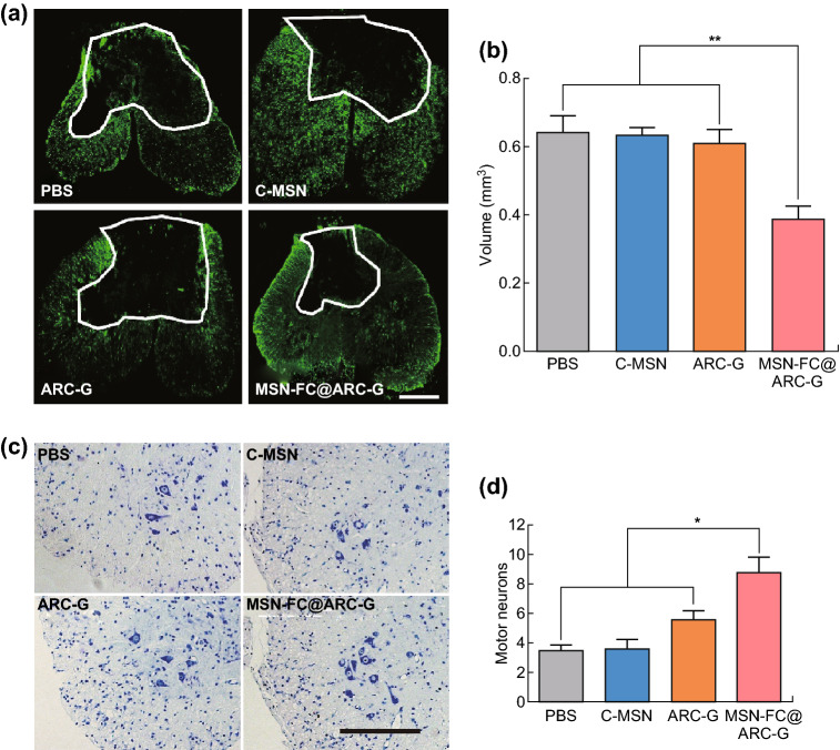
Fig. 7.
a, b Representative injury sites labeled with anti-GFAP antibodies and the statistic histogram of lesion volumes in the four groups (n = 8 mice per group; scale bar: 250 µm). c, d Survival of motor neurons immunostained with Nissl staining in the spinal cord ventral horn (VH) 8 weeks after SCI. (n = 8 mice per group; Scale bar: 250 µm) and one-way ANOVA with Tukey’s multiple comparisons tests; Mean ± SEM; *P < 0.05; **P < 0.01

