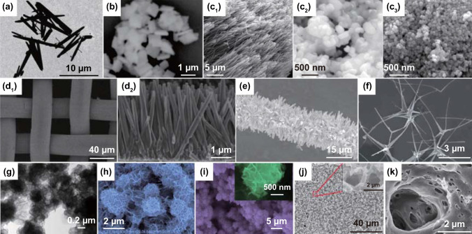
Fig. 5.
a TEM of PZT fibers. Reproduced with permission [67]. Copyright 2014, AIP Publishing LLC. b SEM image of BFO square micro-sheets. Reproduced with permission [70]. Copyright 2017, Elsevier. c SEM images of hydrothermal BTO nanowires, hydrothermal BTO nanoparticles, and commercial BTO nanoparticles. Reproduced with permission [101]. Copyright 2018, Elsevier. d SEM images of bare ZnO nanowire arrays on stainless steel mesh. Reproduced with permission [80]. Copyright 2016, American Chemical Society. e SEM images of ZnO nanowires. Reproduced with permission [88]. Copyright 2015, Elsevier. f SEM image of Ag2O/T-ZnO nanostructures. Reproduced with permission [79]. Copyright 2016, Royal Society of Chemistry. g TEM image of MoS2 nanoflowers. Reproduced with permission [73]. Copyright 2017, Elsevier. h SEM image of WS2 nanoflowers. Reproduced with permission [82]. Copyright 2018, Elsevier. i SEM image of MoSe2 nanoflowers. Reproduced with permission [81]. Copyright 2017, Elsevier. j SEM image of PVDF mesoporous nanostructured film in a top view. Reproduced with permission [83]. Copyright 2014, Elsevier. k PVDF surface image. Reproduced with permission [84]. Copyright 2015, Elsevier. l Micrographs of propidium iodide fluorescent staining cells on cortical bone collagen. The nuclei of the cells are stained in red. The deformed internal side corresponds to the face subject to compression. The deformed external side corresponds to the face subject to tension. Reproduced with permission [85]. Copyright 2017, Trans Tech Publications

