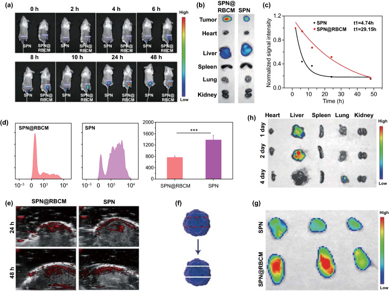
Fig. 4.
a In vivo fluorescence imaging of 4T1 tumor-bearing mice as a function of time after intravenous injection of DiR-loaded SPN or SPN@RBCM. b Ex vivo fluorescence imaging of tumor and major organs harvested after 48 h of intravenous injection. c Normalized fluorescence intensity of SPN or SPN@RBCM in the serum at different time points after intravenous injection. d In vivo uptake of DiO-loaded SPN and SPN@RBCM by Macrophages which was analyzed by FACS. e In vivo PA imaging of 4T1 tumor-bearing mice as a function of time after intravenous injection of SPN or SPN@RBCM. f Schematic illustration of tumor tissue cut into three slices for fluorescence imaging. g Ex vivo fluorescence imaging of tumor slices. h Time-dependent ex vivo fluorescence imaging of major organs

