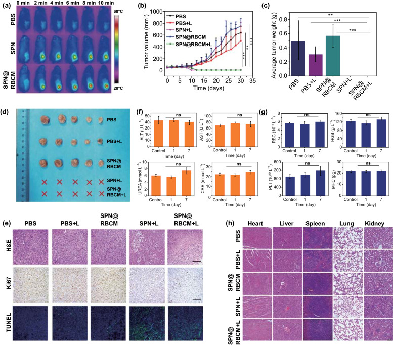
Fig. 5.
a IR thermal images of 4T1 tumor-bearing BALB/c mice under 808 nm laser irradiation (808 nm, 0.5 W cm−2) after PBS, SPN or SPN@RBCM injection. b–d Tumor growth curves (b), average tumor weight (c) and tumor photograph (d) after i.v. injection of PBS, SPN, and SPN@RBCM with or without 808 nm laser irradiation (*p < 0.05, **p < 0.01, and ***p < 0.001; n = 5 per group). e Optical microscopy images of tumor slices stained with H&E, antigen Ki67 and TUNEL after various treatments as indicated above. Scale bar: 100 μm. f, g Blood biochemistry (f) and routine indexes (g) of the BALB/c mice after i.v. injection of SPN@RBCM. h Histological H&E staining of major organs after various treatments as indicated above. Scale bar: 100 μm

