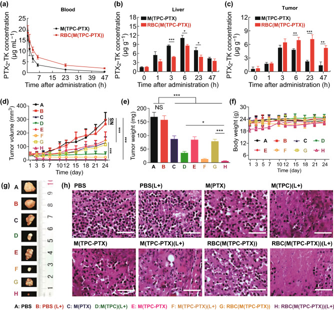
Fig. 4.
a Mice (n = 4) were i.v. administered drug-loaded NPs with or without an RBC membrane coating, using a 15 mg kg−1 PTX equivalent dose. PTX2-TK levels in b the liver and c tumor were assessed at specific time points following NP administration. d Tumor volume changes in mice treated with a range of formulations (PBS, PBS (L+), M(PTX), M(TPC) (L+), M(TPC-PTX), M(TPC-PTX) (L+), RBC(M(TPC-PTX)) and RBC(M(TPC-PTX)) (L+)). L+ indicated laser. e Tumor weights of mice treated as in d. f Body weights of mice over time. g Ex vivo tumor images, as indicated. h Tumor sections were H&E-stained following the indicated treatments. Scale bar = 200 µm. Data d-g are mean ± SEM (n = 6). *p < 0.05, **p <0.01, and ***p <0.001.
Adapted from Ref. [95] with permission

