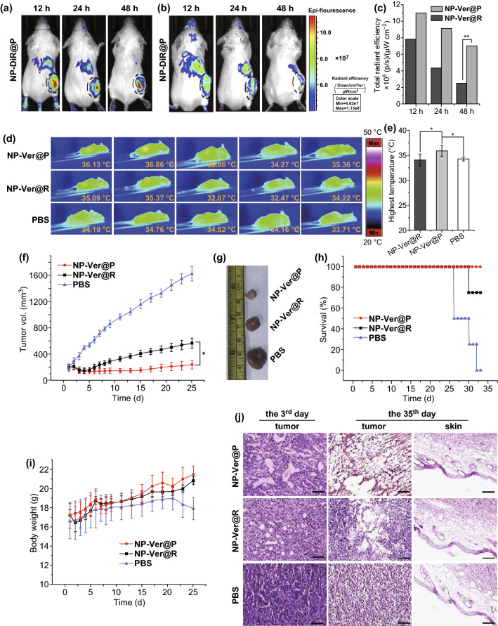
Fig. 7.
Mice received an i.v. injection of NPs, and after 12, 24, and 48 h, tumors and organs of random mice were isolated and used to assess fluorescence and verteporfin levels therein following homogenization. a–c In vivo fluorescence images of mice implanted with 4T1 tumors 12, 24, and 48 h following injection of a PNPs and b RBC-coated NPs loaded with the red membrane dye DiR, with black circled dots identifying tumors. c Total radiant efficiency in tumors was determined based on the images from a and b. d Mice (n = 5/group) were irradiated with 680–730 nm light (0.05 W cm−2) for 10 min, 24 h after administration of PBS or of PNPs or RBC-coated NPs loaded with verteporfin. e Quantification of average tumor center temperatures from d. Data are mean ± S.D. f Average tumor volume in mice following light irradiation. g Tumors were excised from treated mice 35 days following NP administration. h Mouse survival and i average body weight of differently treated mice following light irradiation. Mice treated using RBC-coated NPs loaded with verteporfin are included for reference, as are PBS-injected controls. Data are mean ± standard deviation. j Tumor and skin sections stained with H&E on days 3 and 35 after NP administration. Scale bar = 50 µm. *p < 0.05 and **p < 0.01. Adapted from Ref. [114] with permission

