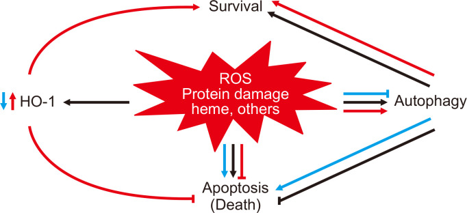
Figure 1. Diagram depicting the relationship between heme oxygenase-1 (HO-1), reactive oxygen species (ROS), and heme in autophagy.
Figure 1 displays subsequent cellular events after cisplatin injury during normal conditions (black arrows), during the absence of HO-1 (blue arrows), and during overexpression of HO-1 (orange arrows). Following cisplatin-induced cell injury (black arrows), there is an attendant upregulation of ROS, which leads to the destabilization of heme-containing proteins and release of toxic free heme. Consequently, autophagy and HO-1 are induced as cytoprotective antioxidant mechanisms within this injurious environment. The lack of HO-1 (blue arrows) worsens cell injury with increased levels of toxic free heme, ROS generation, and oxidative stress, ultimately contributing to dysregulated autophagy and cell death. Conversely, HO-1 overexpression (orange arrows) attenuates cisplatin-induced cell injury by decreasing heme levels, ROS production, and oxidative stress; cells that are better preserved are less in need of autophagy as a cytoprotective response. Reproduced with permission from Bolisetty et al [84].

