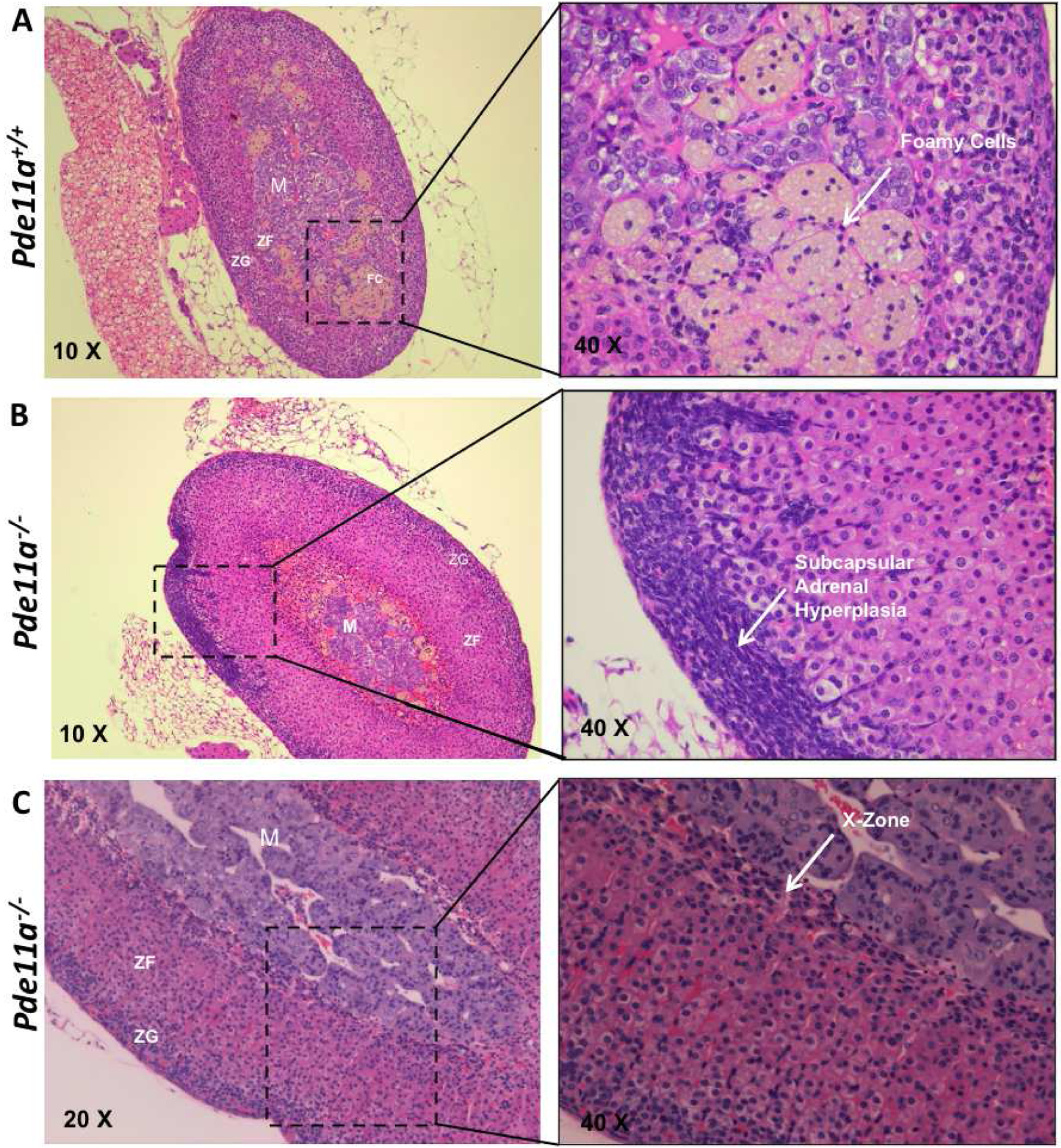
Figure 5: Histological appearance, as shown by H&E staining, of adrenal glands from 12-month old.

(A) Pde11a+/+ and (B & C) Pde11a−/− mice. Arrow point in (A), the presence of foamy cells in the adrenal cortex, more frequently observed in Pde11a+/+; in (B), the presence of subcapsular adrenal hyperplasia in female Pde11a−/− mice; in (C), the persistence of the X- or fetal-zone in female Pde11a−/−mice. Abbreviations: M, medulla; ZF, zona fasiculata; ZG, zona glomerulosa.
