
Figure 6: Persistent X-zone and mRNA expression levels of some markers in wild type (Pde11a+/+) and knockout (Pde11a−/−) animals.
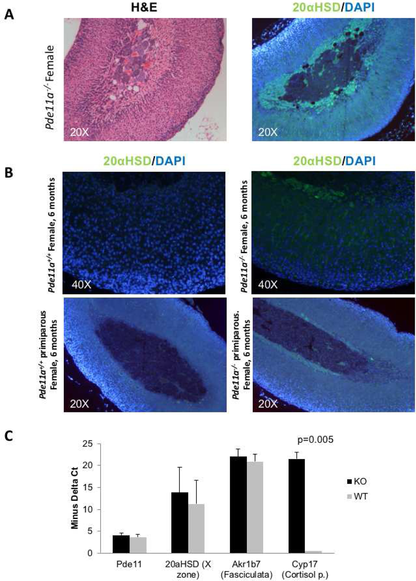
Persistent X-zone as shown by immunostaining with the X-zone-specific marker 20α-hydroxy-steroid dehydrogenase (20αHSD) (green) in Pde11a−/− mice. A. H&E and staining of the corresponding sections with an antibody specific for 20αHSD; B. Presence of the X zone in 2 animals and its absence in their age-matched littermates (one pair at 6 months and the other at 16 months); all are animals that had at least one pregnancy. C. mRNA expression levels of Pde11a, 20αHSD, Akr1b7 and Cyp17 in wild type and Pde11a−/− animals at 6 months of age; Cyp17 is substantially higher in Pde11a−/− animals.