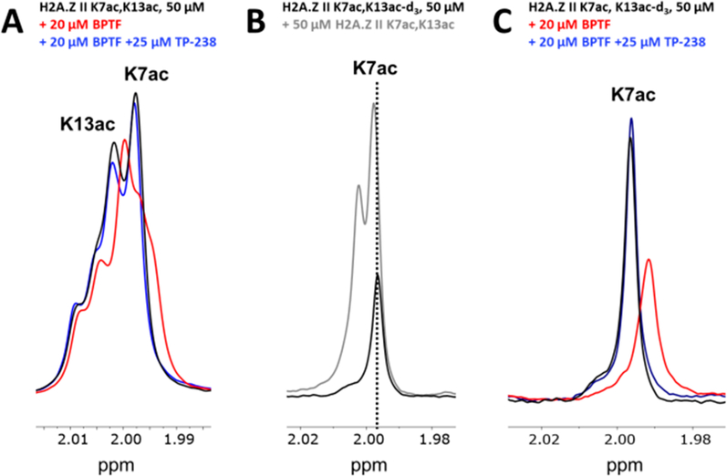
Figure 3.
Ligand-observed 1H NMR CPMG competition experiment evaluating H2A.Z II K7,13ac for BPTF binding site engagement. (A) Experimental spectra of the H2A.Z II K7,13ac peptide alone (black) or with BPTF (red) or competitor TP-238 (blue) are overlaid. (B) Overlaid spectra of the H2A.Z II K7ac,13ac-d3 peptide alone (black) or with the H2A.Z II K7,13 peptide (gray). The dashed line shows the alignment of the K7ac resonance. (C) Experimental spectra for the H2A.Z II K7ac,13ac-d3 peptide alone (black) or with BPTF (red) or the BPTF bromodomain competitor TP-238 (blue) are overlaid.

