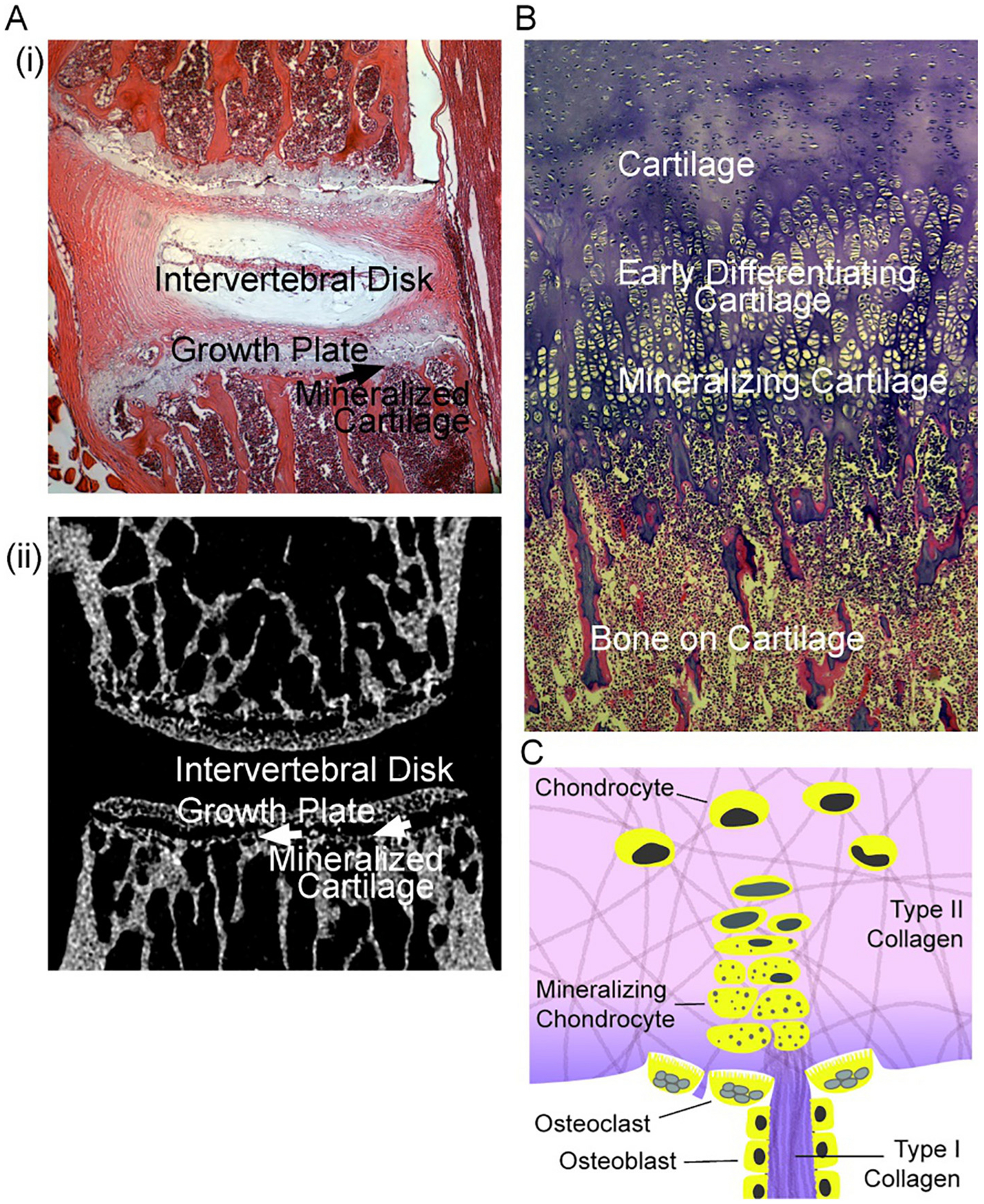
Fig. 2.

Characteristics of mineralizing cartilage. Mouse bone section photographs are unpublished frames of wild type mice [11]; the section of term human bone is an unpublished frame from [30].
A. Left. (i) At the top, a section of mouse vertebrae L2 and L3, showing the disk and adjacent growth plates. (ii) At the bottom, a similar section by micro computed tomography showing the mineralized cartilage at the transition of growth plates to bone, and the adjacent trabecular bone. Each section is 2 mm across.
B. Right. Morphology of cartilage to bone transition in a human term fetus, with cartilage and its transition to bone labeled. This is a section of rib at the transition to bone; in the rib, a broad section of cartilage is involved; narrower regions of growing cartilage occur in vertebral growth plates as in (A) or diagrammed in (C). The region “mineralizing cartilage” is often called “hypertrophic”, see text. The section is 1 mm across and ~2 mm vertically.
D. Diagram of typical structure of a growth plate. Shown are cartilage from the growing plate (top) composed of chondrocytes in mainly type II collagen, and “hypertrophic” chondrocytes that in part lose nuclei and retain vesicles [24] with mineral producing enzymes [31]. At the transition to mineralized cartilage, some type I collagen is present, with trans-differentiation of chondrocytes [32]. Mineralized cartilage (darker color) is degraded mainly by chondroclasts (osteoclasts) and osteoblasts form on this matrix. The authors hypothesize that calcium-phosphate complexes are important in mineralizing collagen, including type I collagen produced in mineralizing cartilage or associated osteoblast-like cells [33], although in early mineralization mineral in vesicles might be important; see text.
