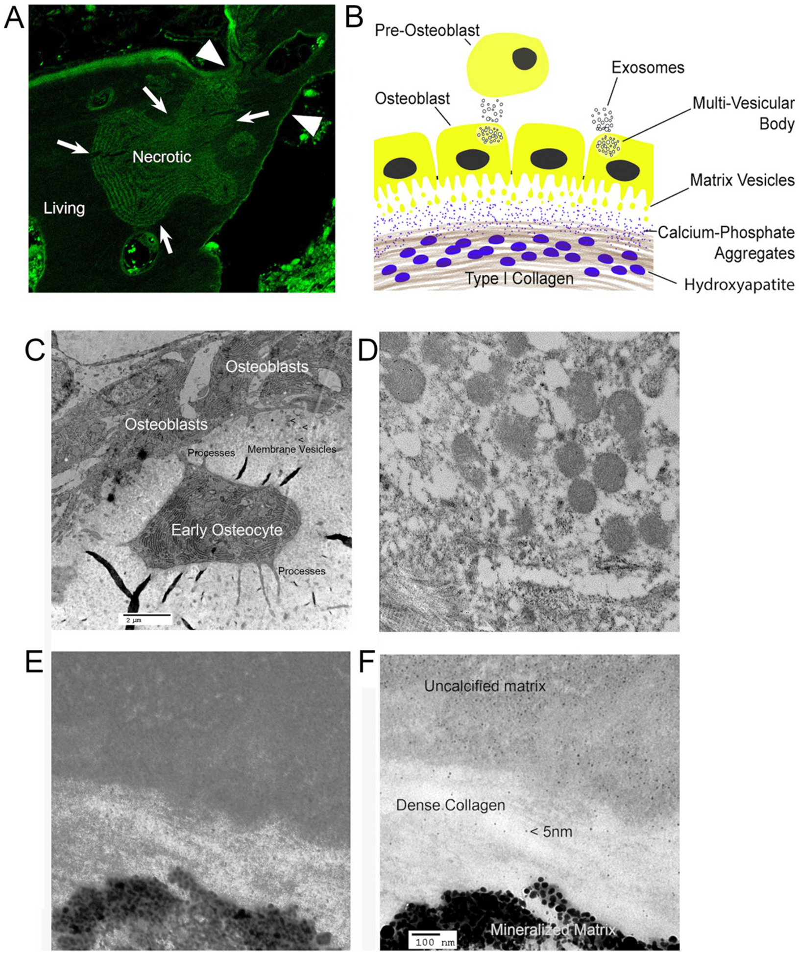
Fig. 5.

Membranes, vesicles, and the apical surface of the bone epithelioid layer. (A) is previously published in [79]; (C–F) are unpublished frames from [40], except (E), from that work, used with permission.
A. Bone is separated from general extracellular fluid by an epithelial-like cell layer. The photograph shows tetracycline labeling of bone formation (top of section) but that tetracycline otherwise is excluded from living (appears dark), but not necrotic (shows labeling) bone; bone is impervious to water and most small ions, but not calcium. For details, see [79].
B. Diagram of bone formation with two types of vesicles. Vesicles, at least in part from multivesicular bodies, are produced on the basolateral surface and serve, at least mainly, to communicate with other cells. At the apical surface, matrix vesicles [80] as in (C) and (D) increase surface area of the cells at the site of mineralization and express phosphate producing enzymes also found in the osteoblast. Matrix vesicles are excluded from dense collagen. Small calcium-phosphate complexes diffuse into dense matrix and form mineral on collagen strands.
C. Plastic embedded thin section of mouse bone in EM (~20 μm across) [40] showing membrane vesicles at the osteoblast apical membrane, the bone secreting side. To retain calcified material, 2 mm thick calvarial bone was fixed 5 min in 4% paraformaldehyde in phosphate buffer at 4 °C and moved directly to dehydration for plastic embedding. An early osteocyte, recently separated from the osteoblast layer, is seen with processes connecting the cell to osteoblasts, and below the osteocyte connecting to deeper osteocytes (not seen here). Irregular black areas are tears in the section.
D. The appearance of this section of the apical surface at higher power. Many vesicles from the membrane are seen. Average vesicle size is ~300 nm. Collagen fibers with crosslinking are seen below the membrane vesicles. Vesicles are excluded from dense collagen fibers, where mineral accumulates (see text).
E. Early mineralized bone on EM of a similar section with dense collagen. Hydroxyapatite crystals ~30 nm associated with collagen are seen. No bilayer vesicles occur in dense crosslinked collagen.
F. The section (E) at higher power. Amorphous calcium phosphates (based on Posner complexes) of small size, ~5 nm, occur in the non-mineralized and dense collagen layer, hypothetical diffusible calcium phosphate aggregates [36,37].
