
Fig. 2.
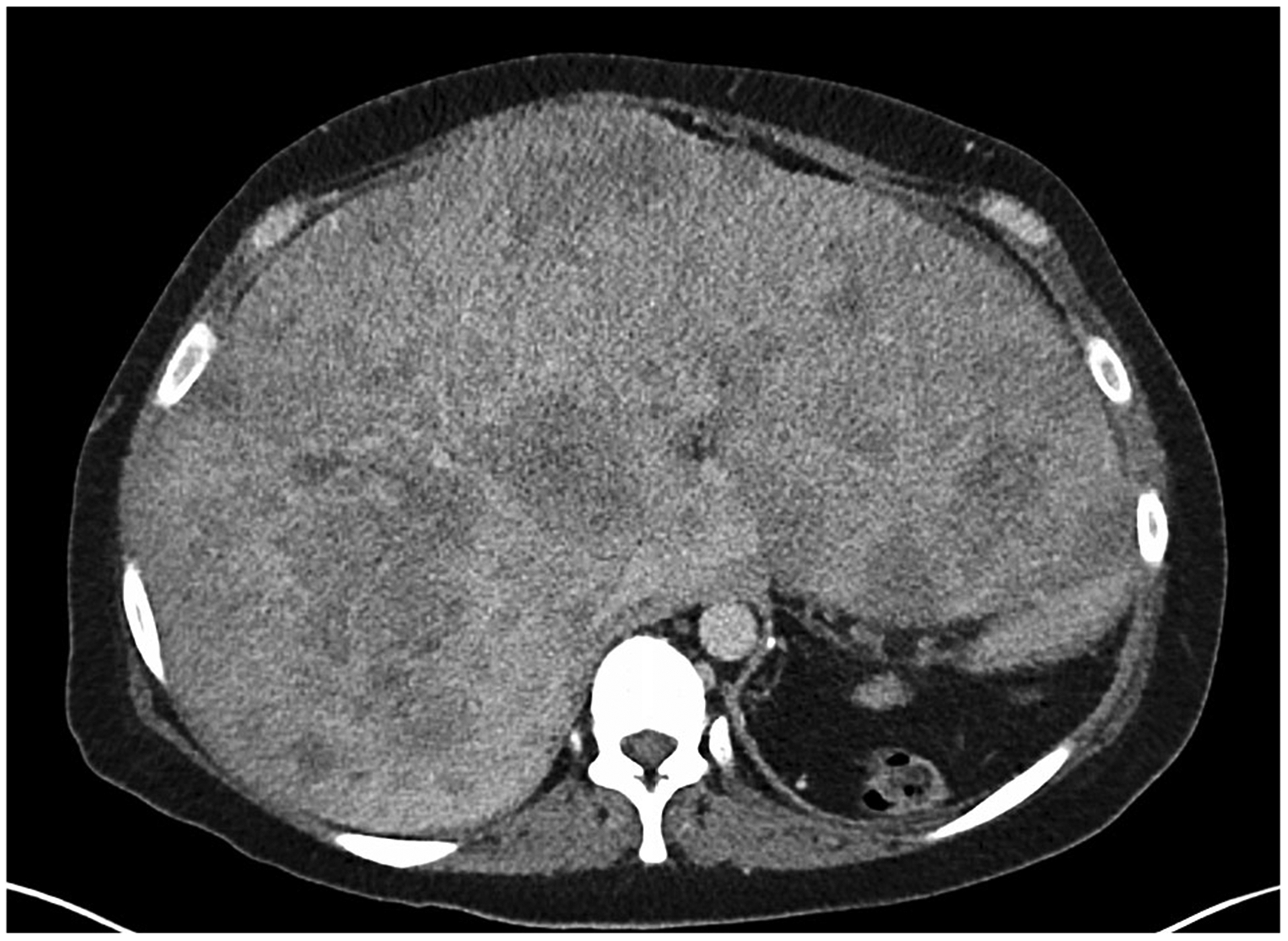
Axial post-contrast CT image from a patient with metastatic breast cancer. The metastases are poorly defined and while they could be measured it is doubtful the measurements would be reproducible from scan to scan and radiologist to radiologist.