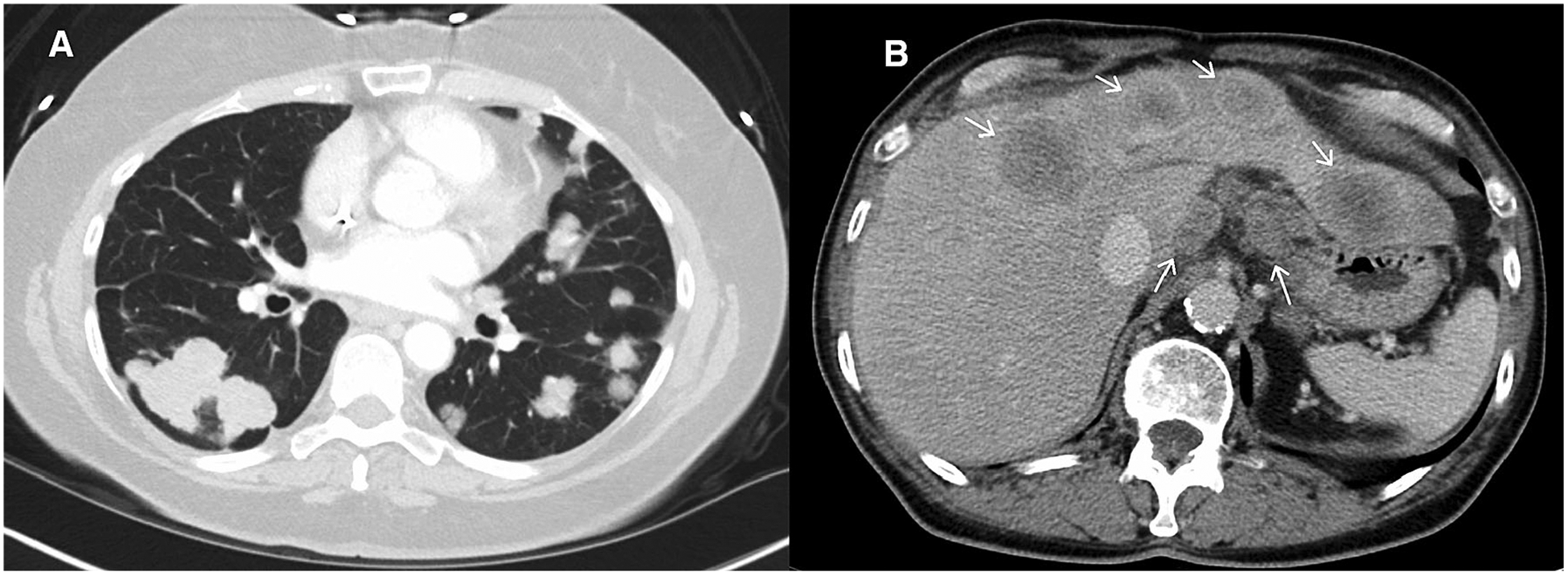
Fig. 5.

Axial post-contrast CT images from a patient with metastatic colon cancer. A shows multiple pulmonary metastases and B shows hepatic metastases and gastrohepatic ligament lymphadenopathy. If these were all accounted for as non-target lesions proper assignment would be: (1) Multiple lung metastases, (2) Hepatic metastases, and (3) Gastrohepatic nodes (assuming short axis measures ≥1 cm).
