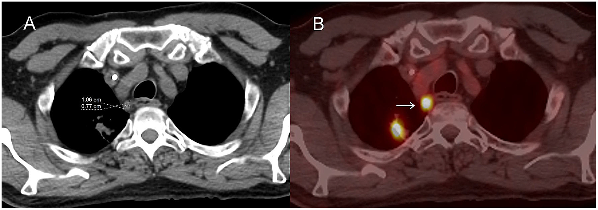
Fig. 6.

Axial images from an FDG PET/CT in a patient with metastatic lung cancer. Non-contrast CT image A shows a 0.8 cm short axis mediastinal lymph node which is hypermetabolic on fused axial PET/CT (B; arrow) and probably a metastatic lymph node. Although this is a site of disease it does not qualify as a non-target lesion because the short-axis is less than 1 cm.
