
Fig. 7.
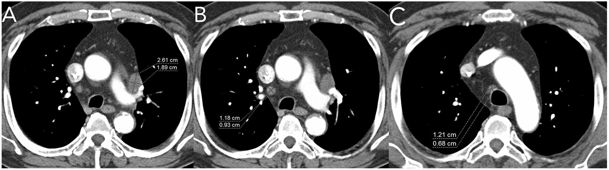
Axial post-contrast CT images from a patient with metastatic lung cancer. A shows a lymph node which was used as a target lesion. A non-target lesion of “other enlarged mediastinal lymph nodes” was also incorrectly delineated for the trial. The other nodes B, C do not meet size criteria for non-target lesions—they measure less than 1cm short-axis.