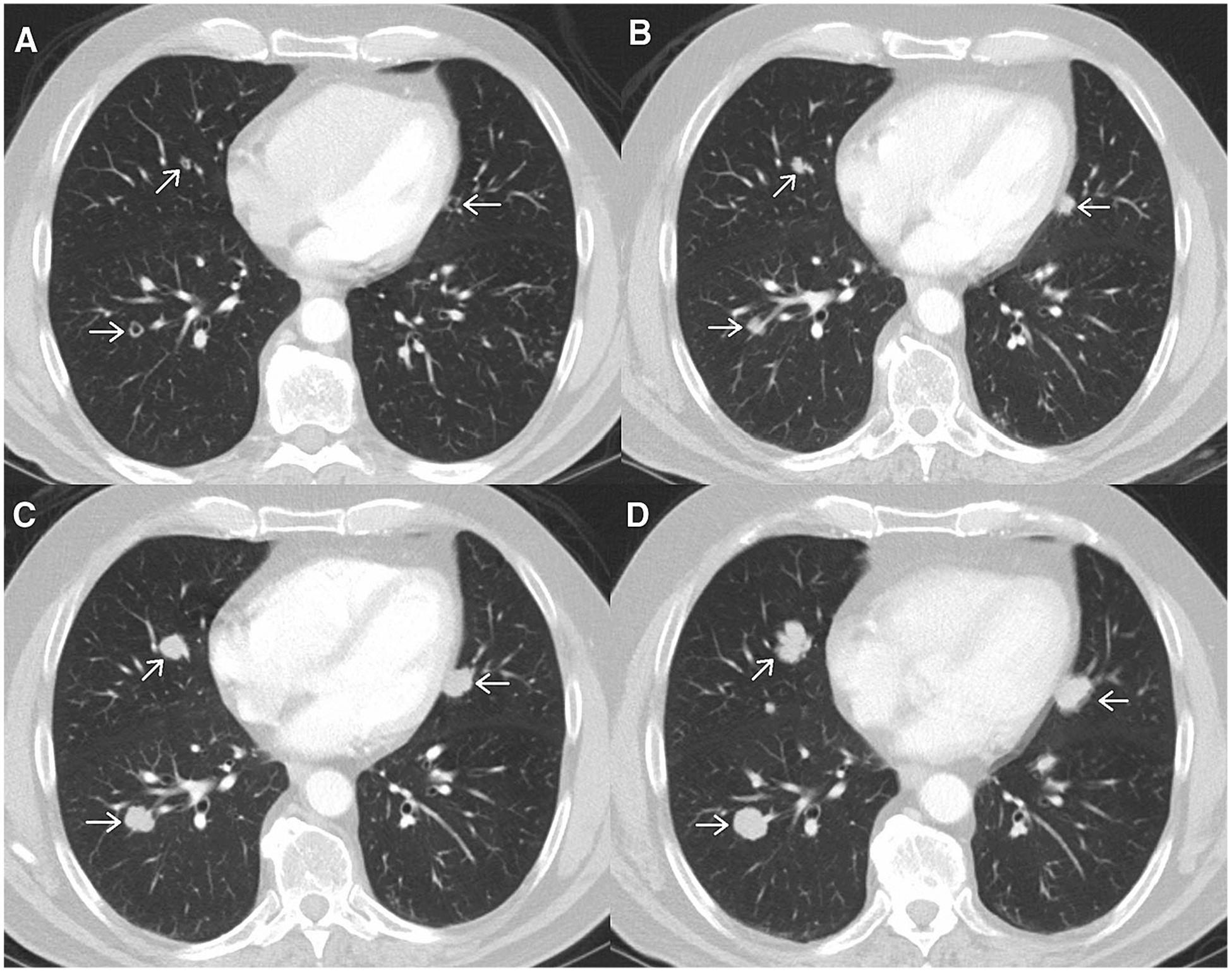
Fig. 8.

Axial post-contrast images from a patient with metastatic melanoma. Initial scan A shows multiple small nodules which can be accounted for as a non-target lesion of “multiple small pulmonary metastases”. Follow-up scans performed at two month intervals B, C, D show gradual increase in size of the nodules. This illustrates a potential difficulty arising when assessing non-target lesions per RECIST v1.1, different radiologists may disagree about the exact time point when “unequivocal progression” occurs.
