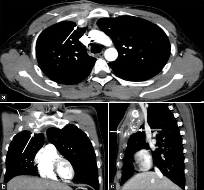
Figure 10:

(a-c) A 43-year-old male presented fever, pain, and localized swelling in the right upper anterior chest wall for 1 month. Contrast-enhanced computed tomography chest (axial, coronal, and sagittal sections in mediastinal window) shows a fairly defined peripherally enhancing collection in the right upper anterior chest wall epicentered in 1st costochondral junction and sternocostal joint (small white arrow). Adjacent encasement of the right internal mammary artery with pseudoaneurysm of the artery is seen (large white arrow).
