
Figure 2:
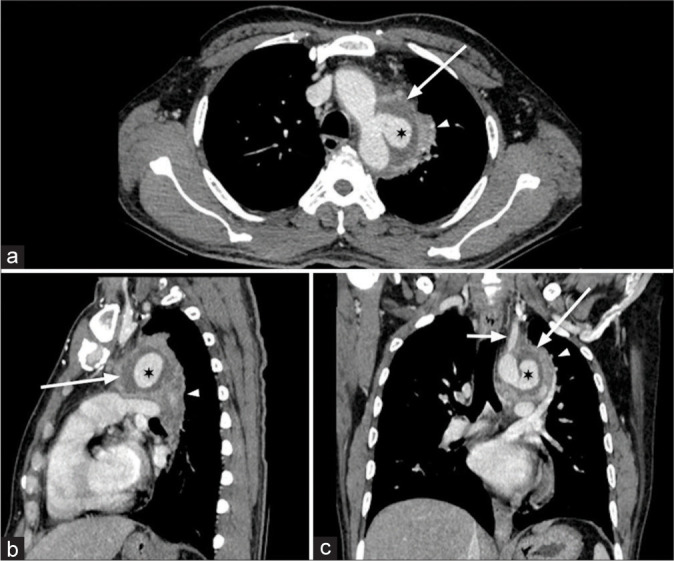
(a-c) A 46-year-old male presented with chest pain for 1 month. Multidetector computed tomography thoracic aortography (axial, sagittal, and coronal sections in mediastinal window) shows focal intense enhancing saccular outpouching (pseudoaneurysm) in the inferolateral wall of aortic isthmus, mushroom shaped (*) just distal to origin of the left subclavian artery (small white arrow) with periaortic hypoattenuating prominent inflammatory soft tissue (large white arrow) causing subsegmental passive collapse of the left upper lobe (white arrowhead).