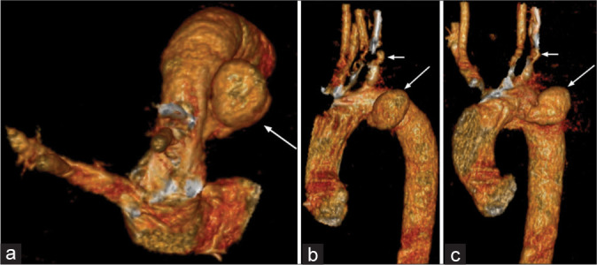
Figure 3:

(a) A 46-year-old male presented with chest pain for 1 month. Multidetector computed tomography (MDCT) thoracic aortography – three-dimensional (3D) volume rendered (VR) image (axial) shows focal saccular outpouching (pseudoaneurysm) in the inferolateral wall of aortic isthmus, mushroom shaped (white arrow) just distal to origin of the left subclavian artery. (b) A 46-year-old male presented with chest pain for 1 month. MDCT thoracic aortography – 3D VR image (sagittal) shows focal saccular outpouching (pseudoaneurysm) in the inferolateral wall of aortic isthmus, mushroom shaped (large white arrow) just distal to origin of the left subclavian artery (small white arrow). (c) A 46-year-old male presented with chest pain for 1 month. MDCT thoracic aortography – 3D multidetector computed tomography image (coronal) shows focal saccular outpouching (pseudoaneurysm) in the inferolateral wall of aortic isthmus, mushroom shaped (large white arrow) just distal to origin of the left subclavian artery (small white arrow).
