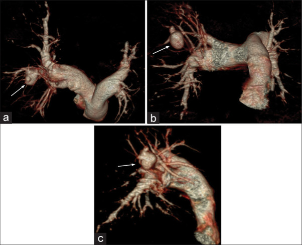
Figure 7:

(a) A 75-year-old male presented with dyspnea, right-sided chest pain, massive hemoptysis, and fever. Multidetector computed tomography (MDCT) pulmonary arteriography – three-dimensional (3D) volume rendered (VR) image (axial) shows focal saccular outpouching (pseudoaneurysm) arising from the feeding posterior segmental branch of the right pulmonary artery (white arrow). (b) A 75-year-old male presented with dyspnea, right-sided chest pain, massive hemoptysis, and fever. MDCT pulmonary arteriography – 3D VR image (coronal) shows focal saccular outpouching (pseudoaneurysm) arising from the feeding posterior segmental branch of the right pulmonary artery (white arrow). (c) A 75-year-old male presented with dyspnea, right-sided chest pain, massive hemoptysis, and fever. MDCT pulmonary arteriography – 3D VR image (sagittal) shows focal saccular outpouching (pseudoaneurysm) arising from the feeding posterior segmental branch of the right pulmonary artery (white arrow).
