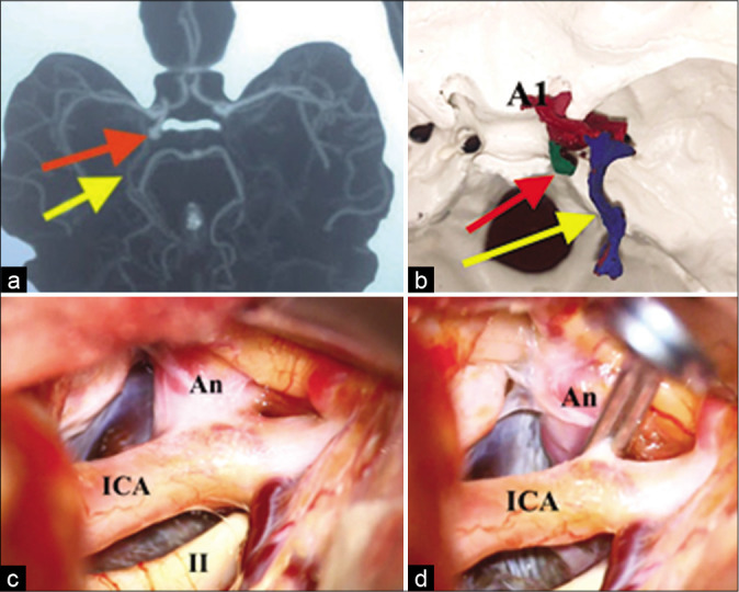
Figure 1:

Images showing the comparison between: (a) the CT angiography image of (5 mm) posterolateral directed right posterior communicating artery aneurysm. The aneurysm (red arrow) and the basal vein of Rosenthal (yellow arrow). (b) 3D physical prototyped biomodel, (c) the intraoperative finding, before the clipping (c) and after the clipping (d). A1: Anterior cerebral artery, precommunicating segment. An: Aneurysm. ICA: Internal carotid artery. II: Optic nerve.
