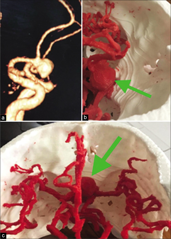
Figure 2:

A case of large anterior communicating artery aneurysm. CT angiography three-dimensional (3D) reconstructed image (a). The 3D prototyped biomodel of the aneurysm, surrounding arteries and the skull base viewed from different angle to enhance preoperative planning and thus intraoperative orientation (b and c). Green arrow: Aneurysm.
