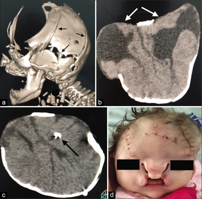
Figure 3:

(a) and (b) Frontal cranial defect with open sutures (black arrows), without craniosynostosis showing two separate sacs with malformed neural tissue (white arrows): dysmorphic ventricle, open lip schizencephaly, lissencephaly spectrum, agenesis of the corpus callosum; (c) ventriculoperitoneal shunt placement (black arrow). (d) After shunt placement.
