
Figure 1: Imaging series from patients.
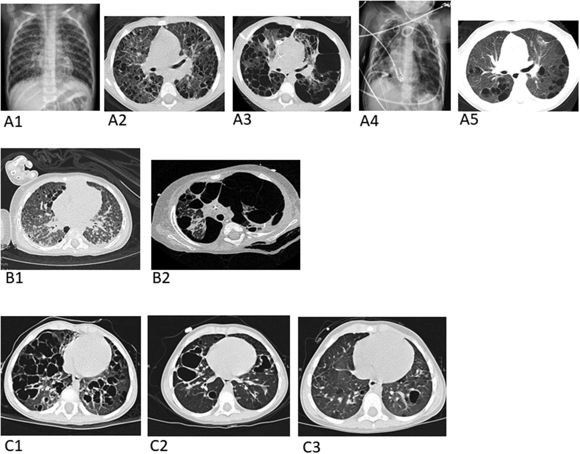
(A) Images from Patient 2: Baseline pre-treatment chest radiograph (A1) and axial chest CT image (A2), depicting multiple pulmonary cysts. An axial chest CT image (A3) one month later shows marked enlargement of many of the cysts with effacement of much of the normal lung parenchyma and bilateral pneumothoraces from cyst rupture. Chest radiograph (A4) three weeks later demonstrates three chest tubes and four pigtailed pleural-drainage catheters placed for refractory air leaks. Axial chest CT image (A5) 15 months later following chemotherapy with cytarabine shows a marked decrease in number and size of pulmonary cysts and reconstitution of normal-appearing intervening lung parenchyma.
(B) Images from Patient 3: Baseline pre-treatment axial chest CT (B1). Axial chest CT (B2) on the day the patient died due to complications of air leaks after 8 weeks of chemotherapy.
(C) Images from Patient 4: Baseline pre-treatment axial chest CT (C1). Axial chest CT(C2) 6 months after starting chemotherapy and axial CT chest (C3) 18 months after starting chemotherapy.
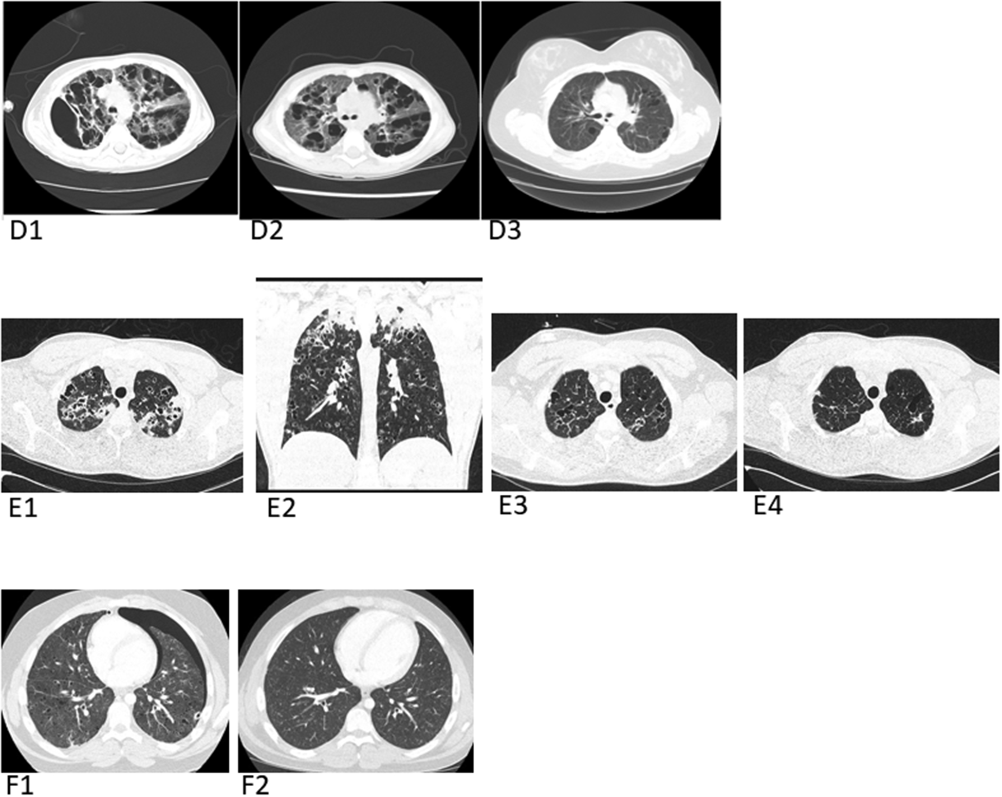
(D) Images from Patient 5: Baseline pre-treatment axial chest CT (D1) shows markedly enlarged pulmonary cysts and axial chest CT (D2) 2 months later shows suboptimal response to vinblastine, prednisone, and methotrexate. Patient was then treated with cytarabine/cladribine combination followed by cladribine alone for 6 months. Post-treatment axial chest CT (D3) several years later shows complete response (CR) of LCH and marked cyst involution with reconstitution of a large percentage of functional lung parenchyma.
(E) Images from Patient 6: Baseline pre-treatment axial chest CT (E1) and coronal chest CT (E2). Axial chest CT (E3) 3 months after starting chemotherapy; Axial chest CT (E4) 10 months after starting chemotherapy.
(F) Images from Patient 7: Baseline pre-treatment axial chest CT (F1) following bilateral chest tube placement for pneumothoraces, reveals a residual left pneumothorax and numerous cysts scattered throughout the lung parenchyma bilaterally. Axial chest CT image (F2) 3 years later off therapy including pleurodesis and chemotherapy, showing resolution of the pneumothoraces and lung cysts.