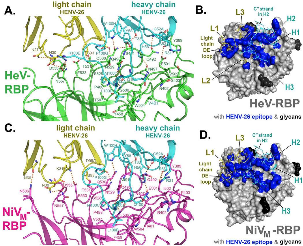
Figure 3. Interface of mAb HENV-26 in complex with HeV-RBP or NiV-RBP.
Residues in the crystal structures of protein complexes with an interatomic distance between antigen (RBP) and antibody of less than 5 Å were designated as participating in the interface.
A) Side view of the interface between mAb HENV-26 and the HeV-RBP head domain. HeV-RBP head domain is colored green, HENV-26 heavy chain in cyan, and light chain in yellow. The interface residues are shown in stick representation. Polar interactions between HeV-RBP and HENV-26 are represented as broken orange lines. The interface residues are labeled in green, cyan, or yellow respectively for HeV-RBP, the heavy chain, or the light chain.
B) Top view of the interface between HENV-26 and HeV-RBP head domain. HeV-RBP head domain is shown in grey as surface representation, glycans on HeV-RBP are colored in black, and interface atoms of HeV-RBP are colored in blue. Interface residues and neighboring residues of HENV-26 are shown as cartoon representation, and interface residues as stick representation. CDRs, the light chain DE loop, and heavy chain C′′ strand of the mAb are labeled.
C) Side view of the interface between HENV-26 and NiV-RBP head domain. NiV-RBP head domain is colored pink, HENV-26 heavy chain in cyan, and light chain in yellow. The interface residues are shown in stick representation. Polar interactions between NiV-RBP and HENV-26 are represented as broken orange lines. The interface residues are labeled in pink, cyan, or yellow for NiV-RBP, the heavy chain, or the light chain, respectively.
D) Top view of the interface between HENV-26 and NiV-RBP head domain. NiV-RBP head domain is shown in grey as surface representation, glycans on NiV-RBP are colored in black, and interface atoms of HeV-RBP are colored in blue. Interface residues and neighboring residues of HENV-26 are shown as cartoon representation, and interface residues as stick representation. CDRs, the light chain DE loop, and heavy chain C′′ strand of the mAb are labeled.

