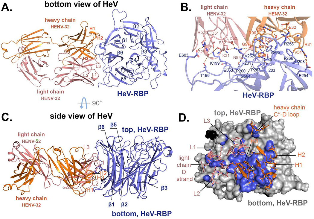
Figure 4. Crystal structure of HENV-32 in complex with HeV-RBP head domain.
A) and C) show cartoon representations of the crystal structures. HeV-RBP head domain is colored in light blue, HENV-32 heavy chain in orange, and the light chain in salmon. Individual CDRs are labeled. The six blades of HeV-RBP head domain are labeled (β1 to β6). Panel A) is the bottom view of HeV-RBP head domain, and panel C) is side view. B) and D) show the interface of the complex (residues with an interatomic distance between antigen and mAb of less than 5 Å). In panel B, interface residues are shown as cartoon representation. The residues of HeV-RBP are labeled in blue, those of the heavy chain in orange, and those of the light chain in salmon. The polar interactions are shown as broken orange lines. In panel D, HeV-RBP head domain is shown as surface representation and colored in grey; the interface atoms from HeV-RBP are colored in light blue. Again, the paratope residues are shown as sticks. CDRs, loop between the heavy chain C′′ strands and D loop, and light chain D strand of the mAb are labeled.

