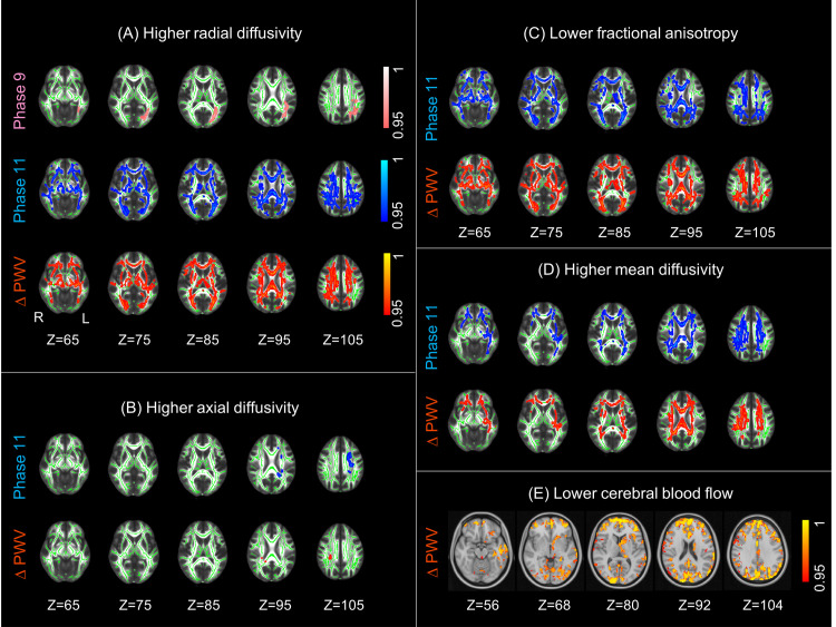
Fig 1. Association of aortic stiffening with WM microstructure and CBF.
Higher aortic stiffening was associated with (A) higher RD, (B) higher AD, (C) lower FA, (D) higher MD, and (E) lower CBF. Associations with Phase 9 PWV, Phase 11 PWV, and ΔPWV are presented in pink, blue, and red yellow, respectively. Five horizontal slices are displayed with MNI152 coordinates ranging from Z = 65 to Z = 105 for WM and Z = 56 to Z = 104 for CBF. The WM clusters are overlaid on the study-specific mean FA skeleton (green) and the standard FMRIB58 FA image, and the CBF clusters are overlaid on the standard MNI152 brain. All results are thresholded at p < 0.05 (TFCE and FWE-corrected p-values) and are presented with a colour gradient for 1 p-values. AD, axial diffusivity; CBF, cerebral blood flow; FA, fractional anisotropy; FWE, family-wise error; L, left; MD, mean diffusivity; PWV, pulse wave velocity; R, right; RD, radial diffusivity; TFCE, threshold-free cluster enhancement; WM, white matter.

