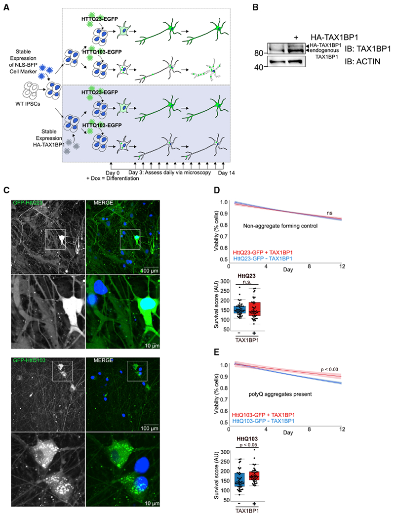
Figure 6. TAX1BP1 Overexpression Preserves Viability in iPSC-Derived Neurons Exposed to Cytotoxic Aggregation-Prone Proteins.

(A) iPSCs were transduced with viruses encoding NLS-BFP, sorted, and transduced with viruses encoding HA-TAX1BP1, followed by puromycin selection. WTiPSCs expressing NLS-BFP with/without HA-TAX1BP1 were transduced with viruses encoding HttQ23-EGFP or HttQ103-EGFP for 24 h, allowed to recover for 24 h, then treated with doxycycline to induce differentiation. Viability was assessed daily for 14 days via imaging of NLS-BFP and quantification of total cell number per well.
(B) Immunoblot confirming TAX1BP1 stable overexpression in iPSCs.
(C) NLS-BFP-expressing WT or TAX1BP1-overexpressing iPSC-derived neurons infected with viruses expressing either HttQ23-EGFP or HttQ103-EGFP.
(D and E) Graphs show line fitted to the number of BFP-positive nuclei counted daily for iPSC-derived neurons with/without stable TAX1BP1 overexpression infected with HttQ23-EGFP (D) or HttQ103-EGFP (E). Ribbon represents 95% confidence interval around the fitted line. Beeswarm boxplots compare survival scores determined by performing permutation analysis using the means of all slopes(center line = median, box limits = first to third quartile, whiskers = minimum and maximum).
