. 2020 Dec 29;9:e59659. doi: 10.7554/eLife.59659

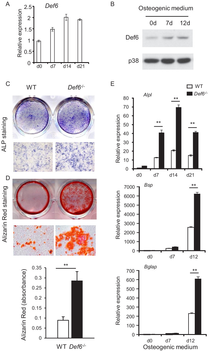
Figure 1. Def6 absence accelerates osteoblast differentiation.

(A, B) Expression of Def6 during osteoblast differentiation using WT and Def6-/- calvarial osteoblast cells at the indicated time points by quantitative real-time PCR (qPCR) analysis of mRNA expression (A) or immunoblot analysis of protein levels (B). (C) ALP staining and (D) Alizarin red staining (upper panel) and its quantification (lower panel) of WT and Def6-/- calvarial osteoblast differentiation at day 15 in osteogenic medium. (E) qPCR analysis of mRNA expression of Alpl, Bsp and Bglap during WT and Def6-/- calvarial osteoblast differentiation process. Data are mean ± SEM. **p<0.01. n.s., not statistically significant.

Figure 1—source data 1. Source data for Figure 1.

Figure 1.

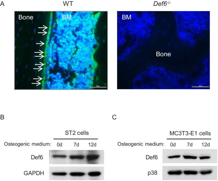
Figure 1—figure supplement 1. Def6 expression in osteoblasts in vivo and in osteoblast cell lines.

Figure 1—figure supplement 1.

(A) Immunofluorescence staining of Def6 (green) on the slices of femurs from 8-week-old WT and Def6-/- mice. Def6-/- mice were used as a negative control for Def6 staining. The slices were counterstained with DAPI (blue). White arrows point to osteoblasts on the bone surface. BM, bone marrow. Scale bar: 50 µm. (B–C) Immunoblot analysis of Def6 expression in ST2 cells (B) and MC3T3-E1 cells (C) during osteoblast differentiation. p38 or GAPDH were used as a loading control.
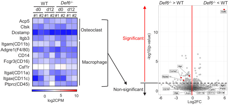
Figure 1—figure supplement 2. RNAseq–based expression heatmap of the marker genes of osteoclasts and macrophages (left panel) and volcano plot (right panel) of RNA-seq analysis of differentially expressed genes in WT and Def6-/- calvarial osteoblasts.

Figure 1—figure supplement 2.

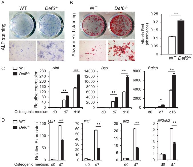
Figure 1—figure supplement 3. CD45 selection does not affect Def6 role in osteoblast differentiation.

Figure 1—figure supplement 3.

CD45 negative calvarial osteoblasts were selected using CD45 magnetic beads according to the manufacturer's instruction, and used to test osteoblast differentiation and gene expression. (A) ALP staining at day 9 and (B) Alizarin red staining at day 21 (left panel) and its quantification (right panel) of CD45 negative WT and Def6-/- calvarial osteoblast differentiation in osteogenic medium. (C, D) qPCR analysis of mRNA expression of osteoblast marker genes Alpl, Bsp and Bglap (C) and ISGs Mx1, Ifit1, Ifit2 and Eif2ak2 (D) during CD45 negative WT and Def6-/- calvarial osteoblast differentiation process. Data are mean ± SD. *p<0.05. **p<0.01.
Figure 1—figure supplement 3—source data 1. Source data for Figure 1—figure supplement 3.
Figure 1—figure supplement 4. Bone marrow derived macrophages (BMMs) from WT and Def6-/- mice were co-cultured with CD45 negative WT calvarial osteoblasts using transwell inserts, in which BMMs were cultured in the upper compartment of the well and osteoblasts were on the bottom compartment of the well.

Figure 1—figure supplement 4.

(A) ALP staining at day 7 and (B) qPCR analysis of osteoblast marker gene expression at the indicated times. n.s., not statistically significant.
Figure 1—figure supplement 4—source data 1. Source data for Figure 1—figure supplement 4.
Figure 1—figure supplement 5. Immunoblot analysis of Def6 expression after TNFα (40 ng/ml) treatment for 24 hr on the WT and Def6-/- calvarial osteoblasts.
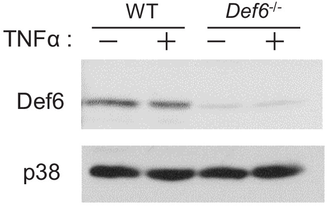
Figure 1—figure supplement 5.

p38 was used as a loading control.