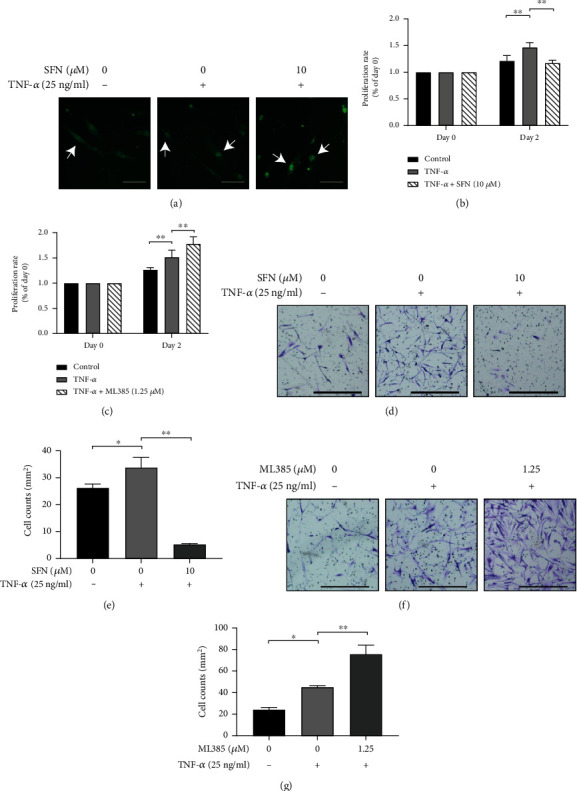
Figure 4.

Effects of nrf2 activator and inhibitor on proliferation and invasion of RA-FLS. (a) Nrf2 nuclear translocation (as shown by white arrows) was tested with immunofluorescence staining in RA-FLS after pretreatment with nrf2 activator sulforaphane (SFN) at a concentration of 10 μM for 1 h and subsequently stimulating with/without TNF-α (25 ng/mL) for 3 h. (b, c) Proliferation of RA-FLS treated with SFN or ML385 was analyzed by CCK-8 assay. Cells seeded in the 96-well plate were pretreated with SFN (10 μM) (b) or ML385 (1.25 μM) (c) for 1 h and subsequently stimulated with/without TNF-α (25 ng/mL) for 2 days. (d–g) Invasion analysis of RA-FLS treated by SFN or ML385 was performed with transwell assay. Cells seeded in the transwell chamber were pretreated with SFN (10 μM) (d, e) or ML385 (1.25 μM) (f, g) for 1 h and subsequently stimulated with/without TNF-α (25 ng/mL) for 2 days. Scale bar represented 200 μm. N = 3. Data were shown as the mean ± SD; ∗p < 0.05 and ∗∗p < 0.01.
