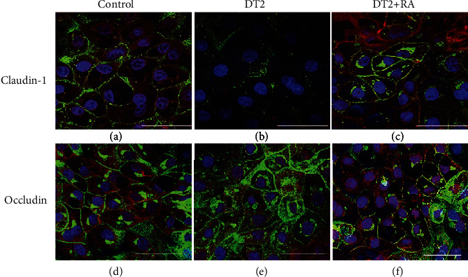
Figure 6.

Effects of DT2 and DT2 + RA (RA pretreatment for 24 h) on the localization pattern of (a–c) claudin-1 and (d–f) occludin using immunofluorescent staining. Differentiated IPEC-J2 cells were cultured on membrane inserts for 10 days then were exposed to DT2 (1 μmol/L DON + 5 nmol/L T − 2) or to DT2 + RA (1 μmol/L DON + 5 nmol/L T − 2 + 50 μmol/L RA); both treatments were applied apically and basolaterally for 72 h. Cells were stained for occludin and claudin-1 (Alexa Fluor 546, red). Cell nuclei were stained with DAPI (blue), and cell membranes were labelled with wheat germ agglutinin (Alexa Fluor 488, green). In controls and in DT2 + RA-treated samples, claudin-1 and occludin were colocalized with wheat germ agglutinin. White scale bar shows 50 μm.
