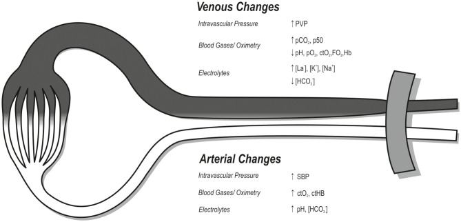
Figure 6.
Illustration of significant changes during BFR compared to regular low-intensity training of the upper extremity (p < 0.05). While the white area reflects the arterial system, the dark area describes the venous system. The tourniquet commonly used in BFR is located proximally, illustrated in gray. Changes are indicated by arrows for increase (↑) or decrease (↓). SBP, systolic blood pressure; PVP, peripheral venous pressure; pCO2, carbon dioxide partial pressure; pO2, oxygen partial pressure; ctO2, oxygen content; FO2Hb, oxyhemoglobin fraction; ctHb, hemoglobin content; p50, oxygen half-saturation pressure of hemoglobin; La−, lactate; K+, potassium; Na+, sodium; , bicarbonate.

