Figure 2.
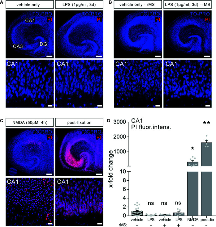
Lipopolysaccharide and/or repetitive magnetic stimulation do not affect cell viability. (A–C) Representative examples of propidium iodide staining (PI, red) in the various experimental conditions (TO-PRO nuclear stain, blue). (A) No effect on membrane integrity is observed following in vitro LPS exposure (1 µg/ml, 3 days) as compared to age- and time-matched vehicle-only treated controls. (B) No signs of cell death are detected 2–4 h after rMS, both in vehicle-only and LPS-treated tissue cultures. (C) NMDA treatment (50 µM, 4 h) and formalin fixation served as positive controls in these experiments. Scale bars: row at the top 200 µm and row at the bottom 20 µm. (D) Summary graph and combined analysis of changes in propidium iodide fluorescence intensity in area CA1 under the indicated experimental conditions, respectively (nvehicle-only = 21 cultures, nLPS = 8 cultures, nrMS = 5 cultures, nLPS+rMS = 6 cultures, nNMDA = 8 cultures, npost-fix = 5 cultures; Kruskal-Wallis test followed by Dunn’s multiple comparisons). Individual data points are indicated by gray dots. Values represent mean ± s.e.m. (* p < 0.05, ** p < 0.01; ns, not significant difference).

