Figure 6.
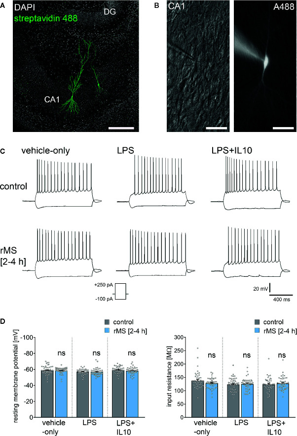
Repetitive magnetic stimulation (rMS) and Lipopolysaccharide have no major impact on intrinsic cellular properties of CA1 pyramidal neurons. (A, B) Examples of CA1 pyramidal neurons recorded and filled with Alexa 488 (10 µM). Cytoarchitecture visualized with DAPI in (A), Dot gradient contrast in (B). Scale bar: (A) 200 µm, (B) 50 µm. (C) Sample traces of input-output properties in CA1 pyramidal neurons from vehicle-only, LPS (1 µg/ml, 3 days) or LPS (1 µg/ml, 3 days) + IL10 (10 ng/ml, 3 days) treated tissue cultures, respectively. (D) Resting membrane potential and input resistance are not significantly different between the groups (vehicle-only: ncontrol = 47 cells in 8 cultures, nrMS [2-4h] = 46 cells in 8 cultures; LPS: ncontrol = 47 cells in 8 cultures, nrMS [2-4h] = 46 cells in 8 cultures; LPS + IL10: ncontrol = 40 cells in 7 cultures, nrMS [2-4h] = 44 cells in 8 cultures; Mann-Whitney test; one recording excluded in the LPS/rMS-group due to high series resistance during recording). Individual data points are indicated by colored dots. Values represent mean ± s.e.m. (ns, not significant difference).

