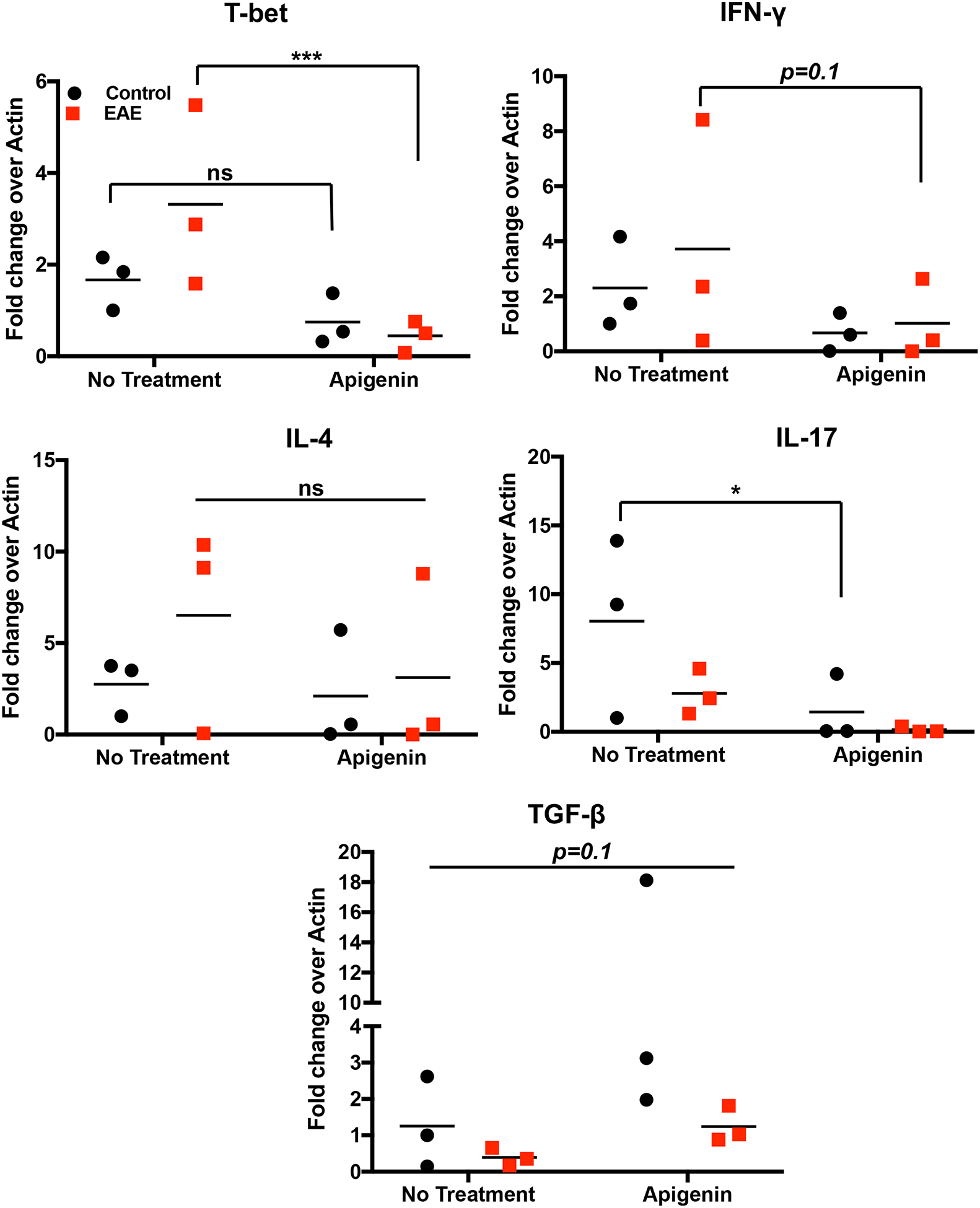
Figure 4. Antigen dependent assay with splenocytes from EAE mice.

Single cell suspension was prepared from mouse spleen of EAE and naïve mice (n=3). Cells were stimulated with and without MOG35–55 peptide for 3 days in the presence of absence of 20μM Apigenin. Gene expression was compared to β-Actin and normalized to cells that were not treated with MOG35–55 peptide. T-bet, IFN-γ, IL-4, IL-17, and TGF-β mRNA expression for three EAE and three naïve mice were detected by qPCR. Statistical significance was determined by 2-way ANOVA using Sidak’s test for multiple comparisons (*p<0.05, ***p<0.001).
