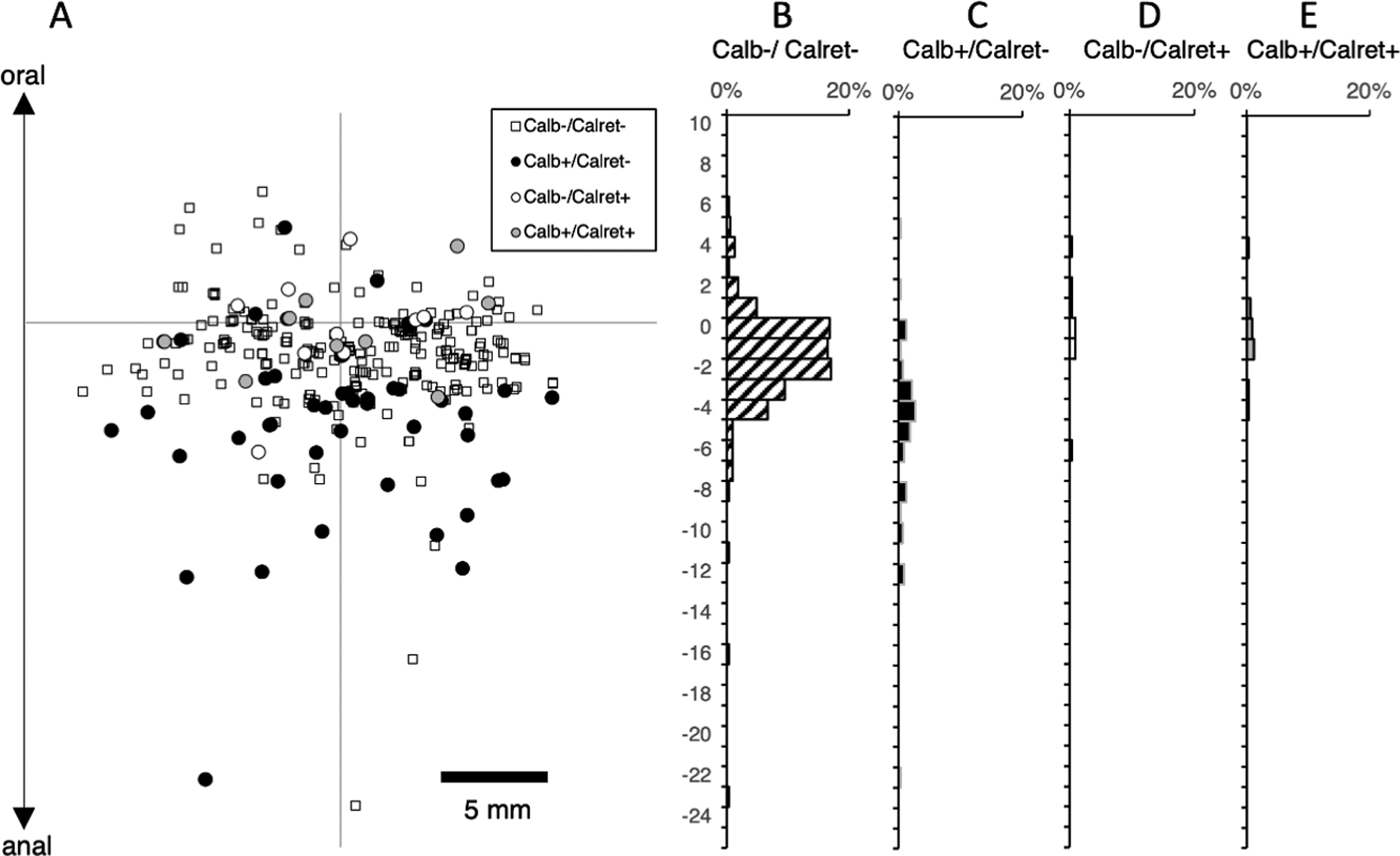
Figure 1:

Ascending motor neurons to the circular muscle, subdivided by calbindin and calretinin immunoreactivities (n=4). DiI was applied to circular muscle near the oral end of the preparation (origin of axes – A) to ensure that ascending projections were preserved; this means that the distribution of neurons with descending projections may have been truncated. Note that there are few motor neurons with ascending projections longer than 12mm; in fact, 95% of all ascending motor neurons lie within 8mm of the DiI application site (origin of the axes). Of ascending motor neurons, 83% lacked both calbindin and calretinin immunoreactivities (B – small square in the scatter plot), 15% were calbindin immunoreactive but few were immunoreactive for calretinin.
