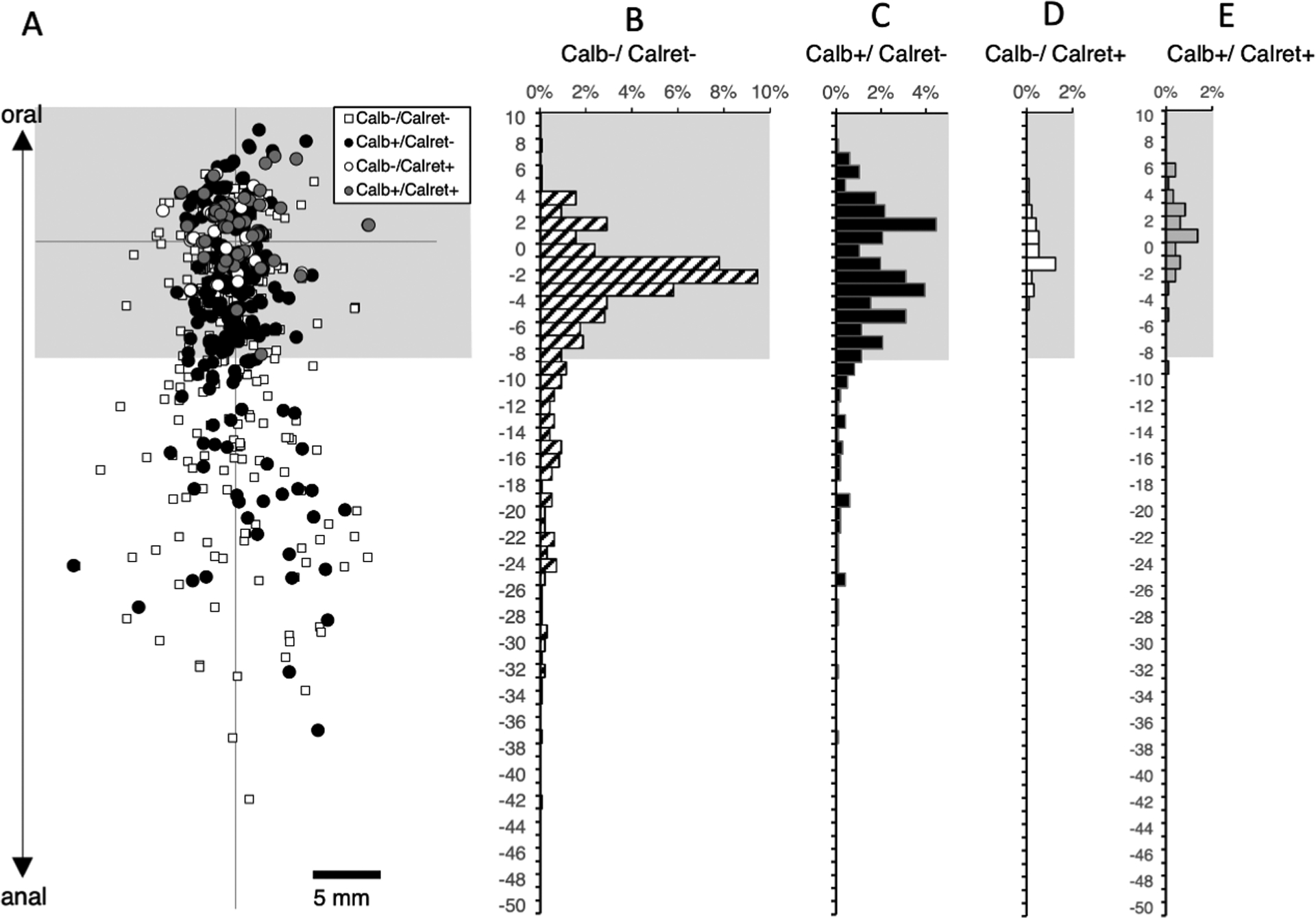
Figure 3:

Ascending interneurons with calbindin and calretinin immunoreactivities. The DiI application site was on the myenteric plexus at the origin of the axes (A). The region where motor neurons were abundant is shown in grey and was excluded from the analysis which concentrated on ascending interneurons located more than 8mm aboral to the application site. Immunohistochemical labelling for calbindin and calretinin was combined with DiI tracing. Calbindin immunoreactive neurons accounted for just under one third of ascending interneurons (32.9% - C). In contrast, calretinin was only present in one cell with a long (>8mm) ascending projection (E) and thus is not a marker for ascending interneurons in the human colon. Most ascending interneurons lacked both calbindin and calretinin (B – small white squares).
