
Figure 4:
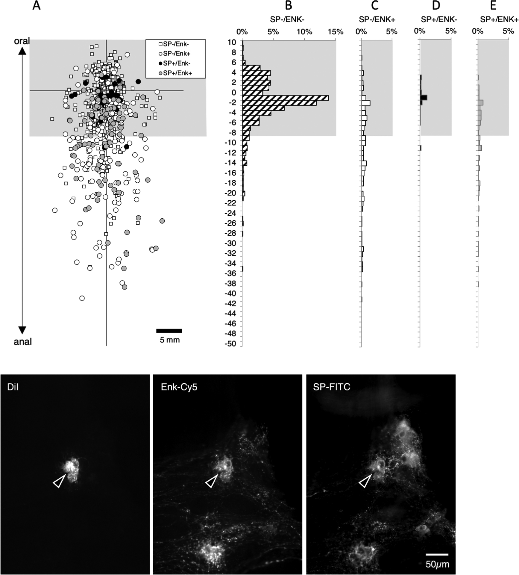
Ascending interneurons labelled with Substance P (SP) and leu-enkephalin (ENK) immunoreactivity. Filled neurons located more than 8mm aboral to the DiI application site on the myenteric plexus (at the origin of axes - A) were considered ascending interneurons (below the greyed area - A). 211 neurons with long ascending projections (>8mm) were labelled: of these 132 (62.6%) were immunoreactive for ENK (C&E) and about one third of the ENK-immunoreactive neurons were also immunoreactive for SP (E). Thus, SP is largely confined to a subset of the ENK-immunoreactive ascending interneurons. Within 8mm of the DiI application site, where motor neuron populations are numerous, there are many neurons with SP which lack ENK (black circles D) and neurons that lacked both ENK and SP (B – small white squares). The micrographs in F show a typical DiI-filled ascending interneuron immunoreactive for both SP and ENK located 14.8mm aboral to the DiI application site.