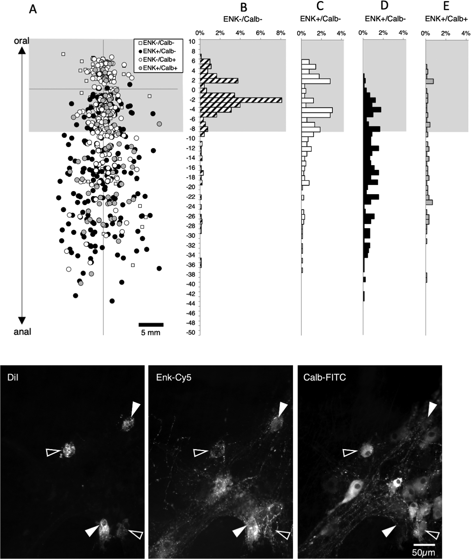
Figure 5:

Ascending interneurons with enkephalin and/or calbindin immunoreactitivies. DiI was applied to the myenteric plexus at the origin of the axes and each symbol (A) represents one DiI-labelled nerve cell body pooled from 6 specimens. Cells within 8mm of the DiI application site (greyed area) were not analysed as these contain a mixture of populations. Histograms (B-E) show the distribution of neurons with immunoreactivity for enkephalin and/or calbindin. Note that in the area occupied by ascending interneurons (below the greyed area), large numbers of neurons were immunoreactive for enkephalin, with or without calbindin (Figure 5D&E); a few cells were immunoreactive for calbindin without enkephalin (Figure 5C and relatively few cells (33 cells of 371; 8.9% of ascending interneurons) lacked both of these markers (Figure 5B – small white squares in A). Micrographs in F show three DiI-filled neurons, located 11.7mm aboral to the DiI application site; two were immunoreactive for calbindin but not ENK (open arrowheads) and two that were immunoreactive for ENK without calbindin (solid arrowheads).
