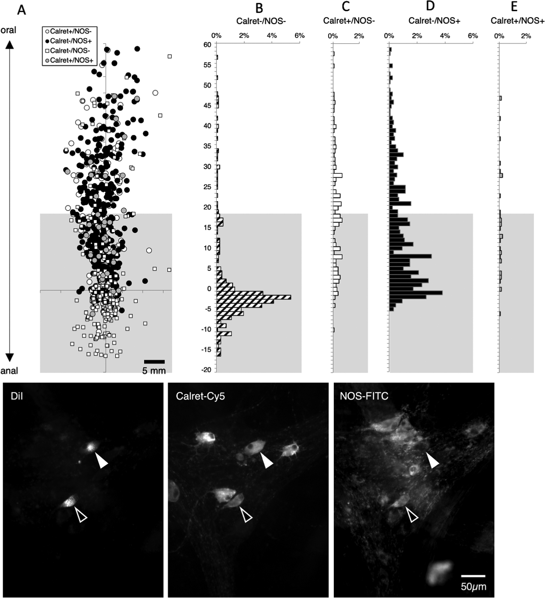
Figure 8:

Descending interneurons – immunoreactivity for calretinin and NOS. DiI was applied to the myenteric plexus, towards the anal end of the preparations and DiI-filled neurons were mapped with their immunohistochemical coding. The greyed area marks where a mixture of motor neurons and interneurons are located (based on lengths of projections); above this is the area more than 18mm oral to the DiI application site where descending interneurons predominate (A). A total of 1288 neurons were labelled with DiI; of these, 329 were located more than 18mm oral to the DiI application site. As expected, the majority of these cells were immunoreactive for NOS without Calretinin (205 of 329, 62.3% - D) or NOS with calretinin (12 cells; 3.6% E). A number of descending interneurons were immunoreactive for calretinin without NOS (78 of 329 cells, 23.7%. C). The remaining 10.3% of interneurons lacked both calretinin and NOS immunoreactivities (B – corresponding to small white squares in A). Micrographs in F show 2 DiI-filled nerve cell bodies located 28.0mm oral to the DiI application site, both of which were immunoreactive for calretinin and one also co-localised NOS immunoreactivity (open arrow).
