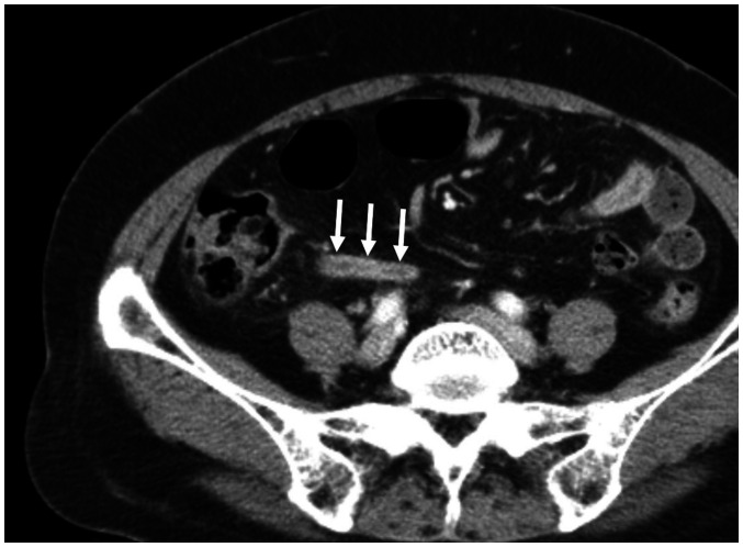
Fig. 4. A 61-year-old woman in the success group.
The contrast-enhanced axial CT shows mild dilatation of the appendix, with a maximal diameter of 8.3 mm, hyperenhancement of the appendiceal wall (arrows), and no periappendiceal fat stranding. She was successfully treated with antibiotic therapy and no recurrence occurred.

