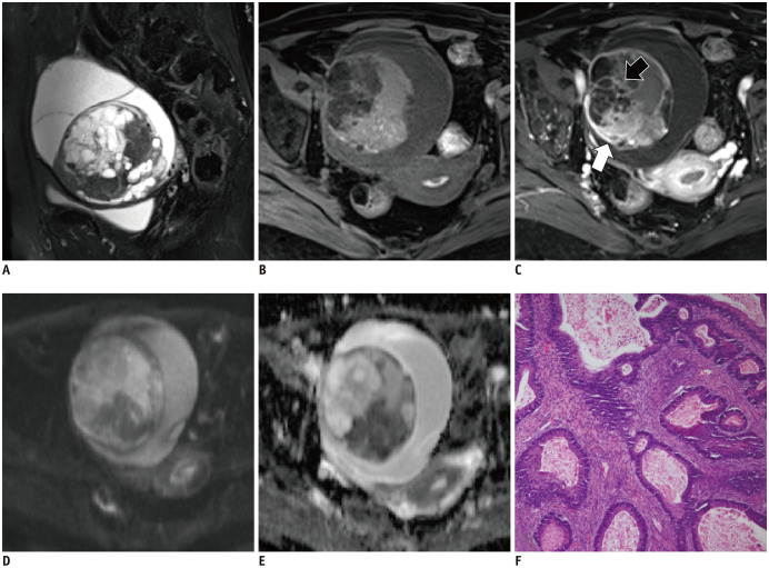
Fig. 3. A 39-year-old woman with right ovarian mucinous borderline tumor.
Sagittal T2WI (A) shows an oval mixed cystic-solid mass with honeycomb loculi. The cystic component exhibited low signal intensity on T1WI (B). The solid component is significantly enhanced (white arrow, C) without restricted diffusion (D, E). The septa within the tumor show irregular thickness (black arrow, C). The corresponding histopathological section (F) shows mild to moderate nuclear atypia, with stratified columnar mucin-containing cells (hematoxylin-eosin staining; magnification × 100). T2WI = T2-weighted imaging

