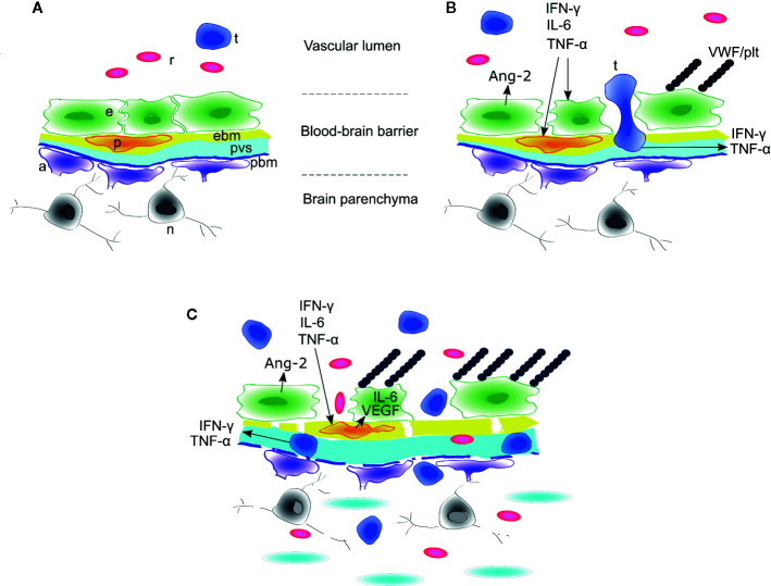
Figure 3.
Model of blood-brain barrier disruption during ICANS. (A) Around cerebral microvessels, the normal blood-brain barrier (BBB) protects the brain parenchyma and neurons by regulating transit of leukocytes and cytokines into the perivascular space. (B) High concentrations of systemic cytokines such as IFNγ, IL-6, and TNFγinduce endothelial cell activation, resulting in release of angiopoietin-2 (Ang-2) and von Willebrand Factor (VWF) from endothelial Weibel-Palade bodies. VWF binds activated endothelium and sequesters platelets. Activated CAR T cells cross the endothelial barrier into the perivascular space and cerebrospinal fluid (CSF). (C) The concentrations of cytokines such as IFNγ and TNFγincrease in CSF due to transit from plasma across the disrupted BBB and/or secretion from CAR T cells or other immune cells undergoing diapedesis. High concentrations of IFNγ and TNF induce secretion of IL-6 and VEGF from brain vascular pericytes and induce pericyte stress, amplifying endothelial activation and BBB disruption. In the most severe cases, this may lead to breakdown of the parenchymal basement membrane and vascular disruption, with thrombotic microangiopathy, acute edema, red blood cell extravasation, microhemorrhage, and neuronal death. a, astrocyte end foot; e, endothelial cell; ebm, endothelial basement membrane; n, neuron; p, pericyte; pbm, parenchymal basement membrane; plt, platelets; pvs,perivascular space and cerebrospinal fluid; r, red blood cell; t, CAR T cell. Figure adapted with permission from (7).

