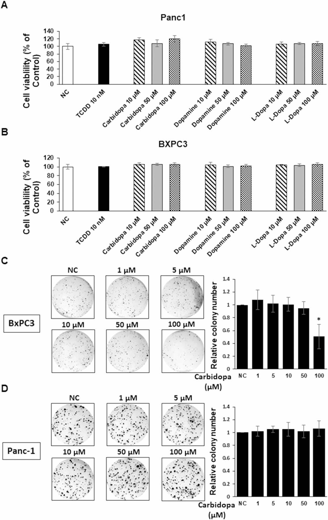
Figure 6.

Cytotoxicity of dopamine, carbidopa, and L-DOPA in Panc1 (A) and BxPC3 (B) cells treated with DMSO, 10 nM TCDD, and 10–100 µM dopamine, carbidopa and L-DOPA for 24 h. Cell viability were determined by cell counting as outlined in the Methods. Cell viability was also determined in BxPC3 (C) and Panc1 (D) cells in a clonogenic assay as described [20] and as outlined in the Methods. Results are expressed as a means ± SD for at least three determinations for each treatment group and significant (P < 0.05) inhibition is indicated (*).
