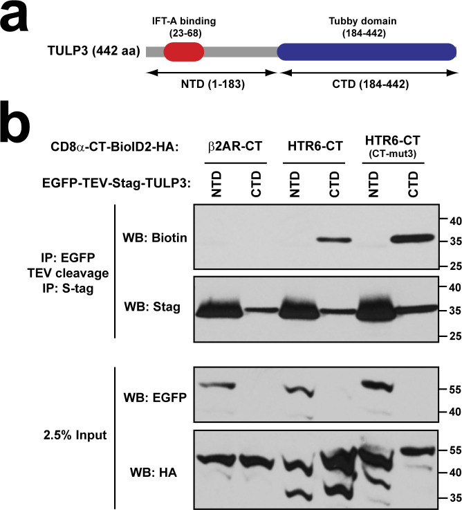
Figure S6. The Tubby domain of TULP3 is responsible for its association to HTR6-CT.
(A) Schematic of TULP3 protein depicting its N-terminal (NTD) and C-terminal (CTD) domains. The NTD contains an IFT-A-binding site, whereas the CTD consists of the phosphoinositide-binding Tubby domain. (B) Proximity biotinylation assay as done in Figs 9 and 10. In this case, biotinylation was analyzed for tandem immunoprecipitated S-tagged TULP3-NTD (aa 1–183) or TULP3-CTD (aa 184–442) (top two panels). Analysis of cleared cell lysates is shown in bottom panels. Molecular weight markers are indicated on the right (kD).

