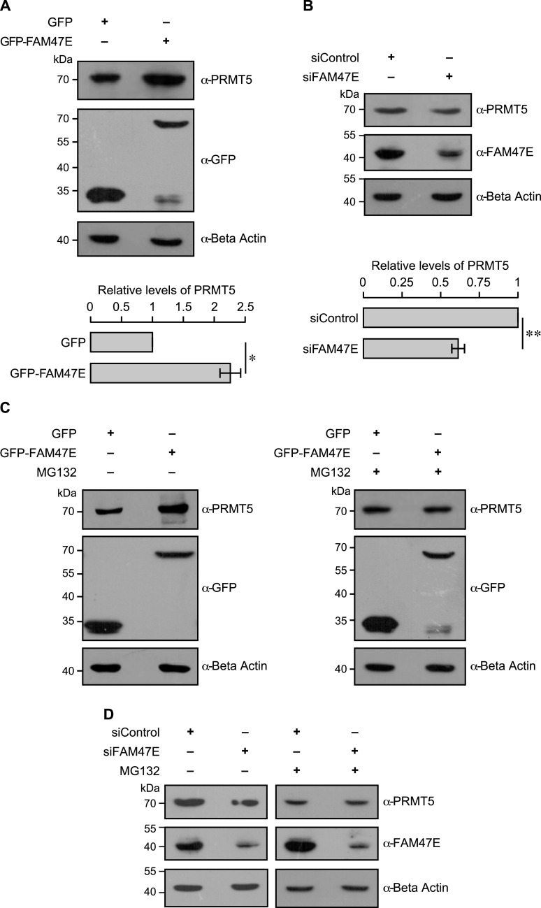
Figure 2. FAM47E increases the stability of PRMT5 protein.
(A) HEK293 cells were transfected with GFP vector or GFP-FAM47E construct. After 48 h of transfection, the cells were lysed, immunoblotted, and probed with PRMT5 antibody or GFP antibody or β actin antibody (upper panel). The band intensities of PRMT5 and β actin in the blots were quantified using ImageJ software and the relative ratios of PRMT5 signal to β actin signal are plotted in the graph (lower panel). The values represent the mean of three independent experiments, with error bars representing SD. Statistical significance was assessed using two-tailed t test. * indicates P < 0.05. (B) HEK293 cells were transfected with control siRNA vector or FAM47E siRNA. After 48 h of transfection, the cells were lysed, immunoblotted, and probed with PRMT5 antibody or FAM47E antibody or β actin antibody (upper panel). The band intensities of PRMT5 and β actin in the blots were quantified using ImageJ software and the relative ratios of PRMT5 signal to β actin signal are plotted in the graph (lower panel). The values represent the mean of three independent experiments, with error bars representing SD. Statistical significance was assessed using two-tailed t test. ** indicates P < 0.01. (C) HEK293 cells were transfected with GFP vector and GFP-FAM47E construct. After 40 h of transfection, the cells were treated with DMSO or MG-132 and incubated for 8 h. The cells were lysed, immunoblotted and probed with PRMT5 antibody or GFP antibody or β actin antibody. (D) HEK293 cells were transfected with control siRNA vector or FAM47E siRNA. After 40 h of transfection, the cells were treated with DMSO or MG-132 and incubated for 8 h. The cells were lysed, immunoblotted, and probed with PRMT5 antibody or FAM47E antibody or β actin antibody.

