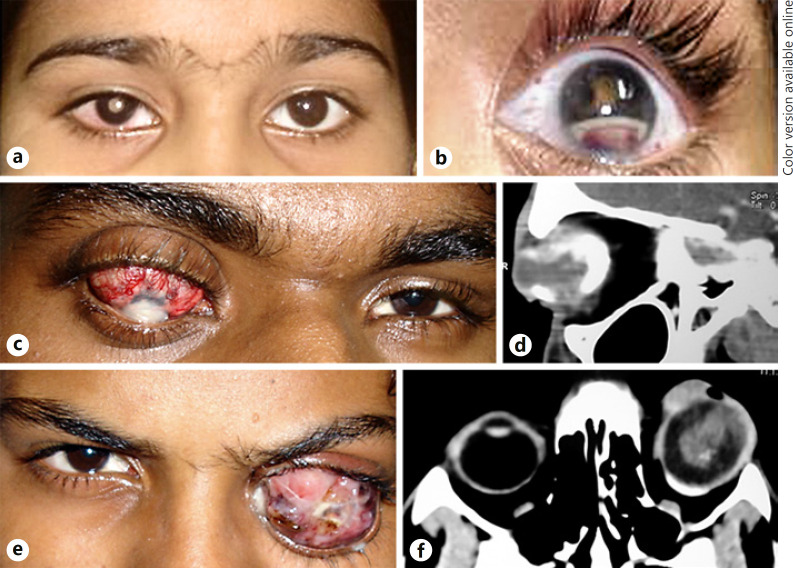
Fig. 1.
a Clinical photograph of a retinoblastoma patient showing leukocoria and circumcorneal congestion of the right eye. b The first symptom noted in this patient was redness, pseudohypopyon, and hyphema in the left eye along with posterior synechiae and leukocoria. This patient had a history of trauma and was misdiagnosed as post-traumatic endophthalmitis. c Clinical photograph of a 17-year-old male patient who presented with staphylomatous eye and corneal opacity following a history of trauma a few years previously. d Contrast-enhanced computed tomographic scan orbit (axial cut) of the same patient showing a calcified intraocular mass within the enlarged globe. e Clinical photograph of a 15-year-old male who presented with proptosis of the left eye following surgery for retinal detachment. f CT orbit scan (axial cut) showing an enhancing intraocular mass with extraocular extension.

